

FIGURE 1.
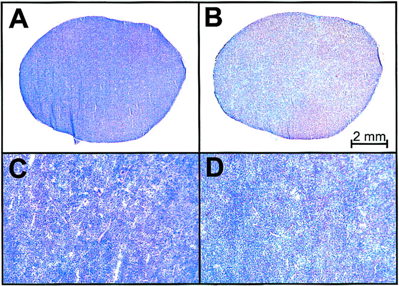
FIGURE 1.
Histologic analysis of spleen obtained from SCID mouse engrafted with primary Ph+ ALL cells. Photomicrographs (×5) of hematoxylin- and eosin-stained splenic section (A) show extensive infiltration of leukemia cells expressing CD19 antigen (B). (C and D) Corresponding sections (×25) demonstrate lack of tissue architecture due to leukemia infiltration. When isotype control antibody was used, no staining was observed (data not shown).


