


Abstract
Gastric emptying in small laboratory animals is a useful parameter to assess gastrointestinal motility for physiologic, pharmacologic, or other research purposes. In mice, phenol red recovery is considered the gold standard for determination of gastric emptying. However, this method requires sacrifice of the animal and yields data of gastric emptying at only 1 time point. Gastric-emptying scintigraphy, the gold standard technique in humans, allows sequential and serial measurements in the same subject. In this study, we developed and validated a novel method of gastric-emptying scintigraphy applied in mice, by comparing it with phenol red photospectrometry. Methods: A dedicated animal pinhole gamma camera was equipped with a specially designed mouse application device. Gastric emptying was measured in unanesthetized mice using pinhole scintigraphy. First, gastric emptying determined with scintigraphy was compared with gastric phenol red recovery simultaneously within the same population. Subsequently, normal values for gastric emptying of solids and liquids in mice were established, and finally, the effects of handling stress and the late effects of frequently used anesthetics or sedatives on gastric emptying were assessed by scintigraphy. Results: Gastric emptying of liquids measured by pinhole scintigraphy did not significantly differ from that measured by phenol red recovery. For the same information, 80% fewer animals were needed for the scintigraphic method. More stress-related delay in gastric emptying was induced by multiple handling of the mice, compared with the less frequent handling that was associated with taking measurements every 10 min or more (P < 0.05). The mean half-emptying time for solids measured by scintigraphy was significantly slower than that for liquid emptying (P < 0.01). Previous anesthesia did not significantly affect gastric emptying 6 h after induction. Conclusion: Dedicated small-animal pinhole gastric-emptying scintigraphy is a reliable tool to investigate gastrointestinal motility in mice, significantly reducing the number of laboratory animals needed for statistical power in trials. The technique enables sequential and serial measurement within 1 subject and is thus useful for follow-up investigations, which can be performed even after invasive procedures that require anesthesia.
Gastric emptying scintigraphy is widely used for clinical and investigative purposes. It is noninvasive, physiologic, easy to perform, safe, and quantitative and is considered the gold standard for the evaluation of gastric emptying of solids and liquids in all types of gastrointestinal disorders. It also allows assessment of the efficacy of gastrokinetic drugs and the effect of surgical procedures on gastric emptying (1–3).
The use of mouse models has increased in gastrointestinal research, particularly in gastrointestinal oncology and gastrointestinal immunology (4). In contrast, gastrointestinal motility research would benefit from further development and application of mouse models (4). In the recent literature, a variety of techniques has been used to assess gastric emptying in small laboratory animals (5–10). In most techniques, the animals are sacrificed, the stomach and intestine are removed and weighed, and the content is analyzed (5,8) by measuring its radioactivity (5), counting the number of glass beads it comprises (9), or assessing it with phenol red photospectrometry (7). Other techniques limit the number of animals needed for an experiment by measuring repetitively without sacrificing the animal, such as by using scintigraphy in a rat parabiosis model (6), MRI (11), or 13C-octanoic acid breath testing in mice (10).
The aims of this study were, first, to adapt a routine pinhole gamma camera system, suitable for murine gastric-emptying scintigraphy without the need for sedation or invasive parabiotic preparation; second, to validate murine gastric-emptying scintigraphy by comparing it with an established technique such as phenol red recovery; and third, to determine normal values for solid and liquid gastric emptying and evaluate the effects of multiple handling, earlier sedation, and analgesia on gastric emptying. Our results indicated that dedicated small-animal pinhole gastric-emptying scintigraphy is a reliable tool to investigate gastrointestinal motility in mice, significantly reducing the number of laboratory animals needed for statistical power in trials and enabling sequential and serial measurement within 1 subject, as is useful for follow-up investigations.
MATERIALS AND METHODS
General Description
Gastric-Emptying Mouse Model.
Female BALB/c mice were purchased from Charles River and maintained under standard conditions at our animal care facility. The mice were used at 8–10 wk old. After an overnight fast, the animals received a test meal and subsequently were scanned without sedation. All animal experiments were performed with the approval of the Ethical Animal Research Committee of the University of Amsterdam and following its guidelines.
Camera Design.
For imaging of gastric emptying in mice, a gamma camera in a dedicated animal care facility was equipped with a pinhole collimator fitted with a 3-mm tungsten insert. The pinhole collimator faced up. On top of the collimator, a specially designed mouse application device was fixed, permitting scintigraphy of anterior projections of mice at a standard orientation and distance from the pinhole aperture without the need for sedation (Fig. 1). The gamma camera was interfaced to a Hermes (Nuclear Diagnostics) acquisition and processing station. Static images of the entire abdomen were obtained for 30 s at the 140-keV 99mTc peak with a 20% window in a 128 × 128 matrix.

Routine pinhole collimator fitted with custom-made plastic adaptor. Using dorsal skin grasp and immobilization against adaptor makes high-quality imaging possible without need for sedation and with standardization of distance and orientation toward pinhole aperture.
Test Meal and Labeling.
Three different test meals were used. The first was a liquid noncaloric test meal that consisted of methylcellulose (OPG Pharma) dissolved in water (30 mg/mL). The solution was labeled with 100 MBq of 99mTc-albumin colloid (Albures; Nycomed-Amersham) per milliliter. For the second test meal, used to compare scintigraphic and photospectrometric assessments of gastric emptying, 0.05% of phenol red (Merck) was added to the 99mTc-albumin colloid-labeled liquid methylcellulose solution. The mice were fed 0.1 mL of the first and second test meals by intragastric gavage. The third test meal, a caloric solid, was prepared by baking 2 mL of egg yolk to which was added 0.5 mL of saline containing 200 MBq 99mTc-albumin colloid per milliliter. The mice voluntarily consumed 100 mg (0.4 kcal) within a few minutes after an overnight fast.
Scintigraphy and Interpretation.
A scintigraphic scan was obtained immediately after gavage feeding (liquid) or voluntary feeding (solid) and every 15 min thereafter (except for the handling stress test) until 60 min. In between scans, the animals were placed back in their cages. Regions of interest (ROIs) were drawn around the total stomach and the intestines, with an additional background ROI drawn on the first postprandial image and copied subsequently on the following images of the sequence (Fig. 2). Meal retention in the stomach was determined as a background-corrected ratio of total abdominal activity. If any gastric emptying was visible on the first image, all counts were considered as gastric content for assessment of the 100% initial value. Total gastric-emptying data were exported to a nonlinear least squares fitting algorithm to determine half-emptying-time (T½) values. The percentage retention of radioactivity at 60 min after the meal (%RRA60) was determined on measured data.
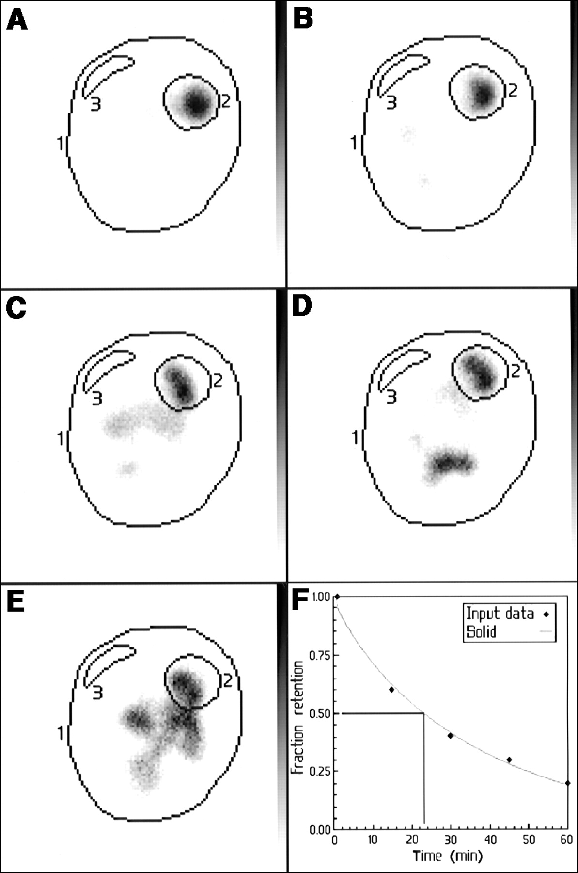
Representative images of gastric emptying in control mouse immediately after solid test meal (A) and every 15 min thereafter up to 60 min (B–E). ROIs are drawn on every acquisition around total field of view (1), stomach (2), and background (3). (F) Gastric-emptying curve derived from measured time points enables assessment of gastric-emptying characteristics and calculation of T½.
Experiments
The experiments had 4 steps:
In the first step, gastric emptying of liquids assessed by pinhole scintigraphy was compared with gastric emptying of phenol red within the same animal population at the same time. Therefore, 25 mice were fed a test meal consisting of 0.1 mL of water with 30 mg of methylcellulose per milliliter and labeled with 99mTc-colloid and 0.05% phenol red dye. The animals were scanned immediately after gavage and subsequently every 15 min up to 1 h. After every scan session, a group of 5 animals was sacrificed and the gastric content of dye was determined with photospectrometry. The animals were killed by cervical dislocation. The stomach and small-intestine fragments (2 cm) were ligated and exteriorized. The tissues were subsequently homogenized for 30 s in 10 mL of a sodium hydroxide concentration of 0.1 mol/L using an Ultra-Turrax (IKA Analysentechnik GmbH). After homogenization, 0.3 mL of 20% trichloroacetic acid was added to precipitate proteins, and the mixture was mixed and centrifuged at 3,000 rpm for 20 min at room temperature. After centrifugation, 2 mL of the supernatant were added to 0.2 mL of a sodium hydroxide concentration of 4 mol/L, and absorbance of the samples was read by photospectrometry at 560 nm. The fraction of gastric retention was calculated according to the following formula: 1 − amount of phenol red recovered at a time point/phenol red recovered at time point 0.
In the second step, the effect of stress by multiple handling on gastric emptying of a caloric solid test meal was assessed. Seven groups of 5 animals were scanned immediately after voluntary ingestion of 100 mg of baked egg yolk and subsequently after varying postprandial time points ranging from once every 5 min to once every 60 min (Table 1).
Scan-Interval Stress Test with Solid Test Meal
In the third step, normal values for gastric emptying of solids and liquids in mice were established. Ten mice received the liquid (oral gavage) test meal after an overnight fast and underwent scanning; 2 wk later, they received the solid (voluntary ingestion) test meal after an overnight fast and underwent scanning. The mice were scanned immediately after gavage or ingestion and subsequently every 15 min up to 1 h.
In the fourth step, late effects of frequently used anesthetics or sedatives on gastric emptying were assessed. Groups of 5 mice were injected intraperitoneally with saline as a control (0.3 mL), diazepam (5 mg/kg; Roche), xylazine (5 mg/kg; Bayer), ketamine and xylazine (100 mg/kg and 5 mg/kg, respectively; Eurovet), or fentanyl/fluanisone (0.3 mL/kg; Janssen Pharmaceutica) and diazepam (5 mg/kg), and 5 mice were anesthetized by inhalation with isoflurane (2.5% for 10 min; Interfarm). All animals were given a liquid test meal by gavage 6 h after induction, followed by gastric-emptying scintigraphy at 15-min intervals.
Statistical Analysis
Differences between 2 groups were analyzed by the nonparametric Mann-Whitney U test. Differences between several independent groups were analyzed by the nonparametric Kruskal-Wallis test. Results are expressed as the mean ± SEM. All statistical tests were 2-tailed, and differences were evaluated at the 5% level of significance. The correlation between gastric-emptying pinhole scintigraphy and phenol red recovery was determined with a nonparametric rank correlation test.
RESULTS
Feasibility
Performing repetitive pinhole gastric-emptying scintigraphy on nonsedated mice was very feasible. Using the small plastic adapter fixed on top of the pinhole collimator ensured identical distance and orientation toward the pinhole aperture. Furthermore, we easily immobilized the animals by grasping their skin and holding them against the adapter without squeezing (Fig. 1). If the technique is performed correctly, the animal will not move or protest and a high-quality acquisition of 30 s can be made without any problem.
An example of a typical pinhole gastric-emptying scintigram in mice is shown in Figure 2. Gastric emptying of a solid test meal is clearly assessable on sequential images and the subsequently derived gastric-emptying curve. The stomach was easily delineated on the first postprandial images. Viewing the image sequence and copying the ROIs on subsequent images were not difficult.
Comparison of Phenol Red and Scintigraphy
The results of the first experiment are shown in Figure 3A. There was no significant difference between the gastric emptying of liquids measured by pinhole scintigraphy and the gastric emptying of liquids measured by phenol red photospectrometry. The T½ values for scintigraphy and photospectrometry calculated from the least squares exponential fit of the mean individual time-point values were 13.3 and 14.5 min, respectively. Scintigraphic quantitation of gastric emptying was accurate: There was a good correlation (linear regression slope: R = 0.96, R2 = 0.93, r = 0.86, P < 0.001) of individual time-point measurements between gastric retention determined by scintigraphy and gastric retention determined by photospectrometry (Fig. 3B). With the scintigraphic method, 80% fewer animals could be used to obtain the same information (phenol red recovery, n = 25; scintigraphy, n = 5).
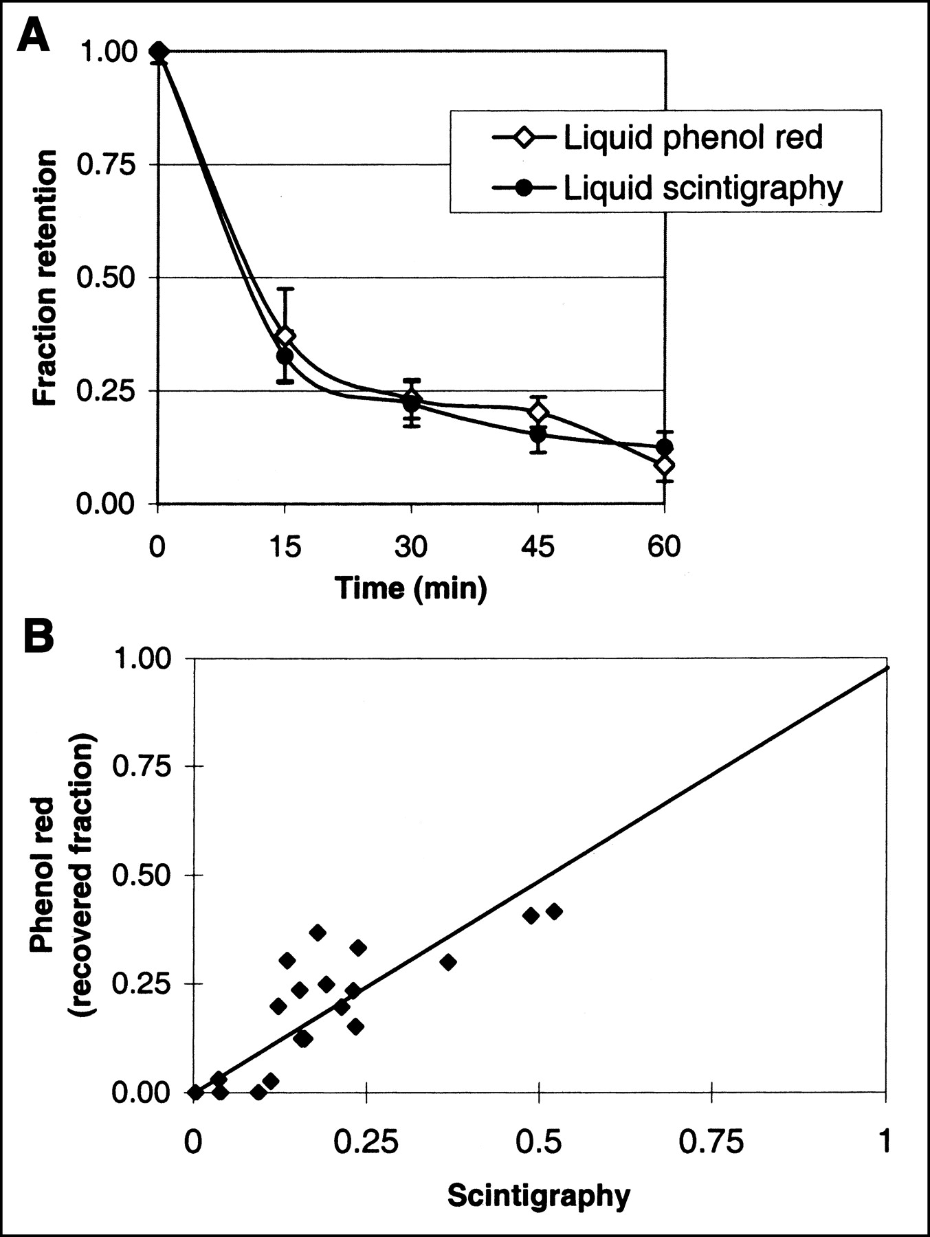
(A) Gastric-emptying curves of liquid test meal labeled with phenol red and 99mTc-albumin colloid. Gastric emptying measured with phenol red photospectrometry (liquid phenol red) did not significantly differ from gastric emptying measured with pinhole scintigraphy (liquid scintigraphy). (B) Individual time-point measurements of gastric phenol red recovery and gastric retention measured with pinhole scintigraphy. Correlation between methods was good (R = 0.96; r = 0.86; P < 0.001).
Effect of Handling
The results of the second experiment are shown in Table 1. The effect of multiple handlings on animals is clearly demonstrated. Both T½ and the residual radioactivity measured 60 min after the meal (%RRA60) were significantly larger in animals that were frequently handled, that is, every 5 or 7.5 min (P < 0.05). At intervals of ≥10 min, T½ and %RRA60 remained stable, indicating that multiple handlings should be avoided. The T½ for group 7 was not calculated because the curve-fitting algorithm needed a minimum of 3 measured input values. Based on these observations, we decided to proceed with 15-min-interval scanning for the remaining experiments, minimizing stress-related interference and yielding enough measurement points for data processing.
Liquids Versus Solids
The results of the third experiment, determining normal values for solid and liquid gastric emptying, are shown in Figure 4. Gastric emptying was significantly slower for solids than for liquids (P < 0.01). The T½ values for solids and liquids were significantly different (28.41 ± 1.51 min and 14.81 ± 2.13 min, respectively, P < 0.001). The normal ranges (mean ± 2 SDs) for solids and liquids were 18.85–37.97 min and 10.57–19.05 min, respectively. The %RRA60 was significantly higher for solids than for liquids (25.45% ± 2.85% and 12.12% ± 2.87%, respectively, P < 0.01). The upper normal values (mean + 2 SDs) for solids and liquids were 34.36% and 21.20%, respectively.

Gastric-emptying curves of solid and liquid test meals (mean ± SD). Solid gastric emptying was significantly slower than liquid emptying (P < 0.01).
Effects of Anesthetics
The results of the final experiment, assessing the effects of previous anesthesia on gastric emptying, are shown in Table 2. All animals survived anesthesia and were well awake 6 h after induction. Previous anesthesia had no significant effect on gastric emptying 6 h after induction, as assessed by T½ or %RRA60.
Gastric Emptying 6 Hours After Anesthesia
DISCUSSION
The data presented here show that gastric-emptying scintigraphy in small laboratory animals is feasible and accurate and yields T½ values comparable to those obtained with the gold standard, phenol red. However, gastric-emptying scintigraphy significantly reduces the number of mice needed and allows temporal measurements on the same animal. These major advantages over invasive techniques make gastric-emptying scintigraphy a reliable tool in gastrointestinal motility research.
Gastric-emptying scintigraphy with radiolabeled standard meals is regarded as noninvasive, physiologic, easy to perform, safe, and quantitative and has become the gold standard for the evaluation of gastric emptying of solids and liquids in all types of gastrointestinal disorders and for assessing the efficacy of gastrokinetic drugs and surgical procedures in humans (1–3,12). Despite the expanding use of mouse models in general, many areas of gastrointestinal research have not taken advantage of the potential of the mouse. For instance, studies on gastrointestinal motility would benefit from further development and application of mouse models, with adaptation of successful methodologies of large-animal physiology to the more minute scale of the mouse (4).
Improvement of scintigraphic resolution, enabling imaging of small laboratory animals, can be achieved using a pinhole collimator, both for planar imaging and for SPECT (13–15). Pinhole collimators are routinely available in most nuclear medicine facilities. To obtain magnified images with high spatial resolution in small-animal studies, system sensitivity is sacrificed and more radioactivity or a longer acquisition time is needed. In mice, increasing the amount of radioactivity administered relative to body weight (compared with humans at a factor of 1,000) results in statistically useful pinhole images with a 7-fold magnification that limits the radiation burden to operators to no more than 10 MBq at any moment. The stomach is easily delineated on the first postprandial images, when, in almost all the animals, the entire test meal is within the stomach. When a liquid test meal is used, some gastric emptying might have occurred when the first postprandial images are obtained. In that case, for quantification, all activity is considered to be within the stomach on the first postprandial images.
Two types of test meal were used. The liquid meal containing methylcellulose to increase viscosity and slow gastric emptying is suitable for mixing with 99mTc-albumin colloid and phenol red (7). The solid meal consisting of baked egg yolk mixed with 99mTc-albumin colloid is known for its stable binding to the solid component (1). The advantage of a liquid meal is the possibility of oral gavage, standardizing the timing of meal administration. The disadvantage is the added stress to the mouse from being handled during oral gavage and the technical difficulty of the procedure, with the possibility that pulmonary instillation or fur contamination will occur, leading to dropouts. The advantage of a solid meal is the reduced stress to the mouse. In our experience, healthy mice eat well after overnight fasting. Timing, however, can be more problematic when one is scanning larger series of animals in a single session, since mice will not always eat immediately on command. Anesthetized or surgically treated animals might not be so eager to eat, possibly complicating logistics in a trial. Depending on the specific trial design, one could choose a solid or a liquid test meal optimizing research circumstances.
Stress has a major impact on gastrointestinal motility, and animal handling should therefore be minimized. To evaluate the effect of handling, we assessed gastric emptying using an increasing number of acquisitions per 60-min scan session. The test was performed with a solid meal to prevent the stress that would be induced by oral gavage of liquids. Our data clearly showed a difference in gastric emptying, with significant slower T½ values when mice were handled more than once every 10 min. Gastric emptying did not significantly differ between the groups when the mice were handled less frequently than once every 10 min, suggesting that the stress level created by this frequency is acceptable and does not cause greater interference with gastric emptying than when the animals were handled once or twice. These observations are in concordance with recently published physiologic-stressor data describing handling as a moderate stress inducer, with rapid normalization of stress-related neurotransmitters after cessation of handling (16). Further, the delay in gastric emptying observed in our experiment agrees with literature reporting that delayed gastric emptying and accelerated large-bowel transit are the most consistent pattern of gastrointestinal motor alteration induced by acute stressors (17). To minimize handling-related changes in gastric emptying, we decided to reduce acquisitions to once every 15 min for 1 h, resulting in enough time points for line-fitting algorithms.
When comparing gastric-emptying scintigraphy in mice to the frequently used phenol red test, which can be considered a historic gold standard (18), we preferred working with a liquid test meal for combining 99mTc-albumin colloid and phenol red. The good correlation between individual measurements proves that gastric emptying can be assessed accurately with pinhole scintigraphy. Eighty percent fewer animals were required for gastric-emptying pinhole scintigraphy than for phenol red recovery to yield the same information. When animals do not need to be sacrificed for other invasive testing, repeated scintigraphic studies could be performed for longitudinal studies, even further decreasing the number of animals needed.
When pinhole gastric-emptying scintigraphy is compared with the recently introduced noninvasive 13C-octanoic acid breath test (10,19), both tests are seen to be noninvasive and to need no animal sedation. Both tests can be repeated on the same subjects since the animals do not need to be sacrificed for information gathering. The slight increase in the frequency of animal handling for scintigraphy, compared with breath testing, seems not an interfering factor, as long as acquisition is limited to once every 15 min. Scintigraphy requires a radiopharmaceutical, resulting in a limited radiation burden to the animal handlers. Breath testing with stable isotopes does not have this disadvantage. Scintigraphy, however, offers quantitative as well as visual information on gastric emptying, compared with the only-quantitative information offered by breath testing. Supplemental visual information might eventually help the individual outlier results of experiments to be better understood (10). Both tests require specific instrumentation not always routinely available in every research facility.
As in humans, a difference in gastric emptying between solids and liquids is expected (20). In many gastric-emptying studies on mice, the viscosity of the liquid test meal is increased to slow emptying by adding methylcellulose (7,19). The series of normal values for methylcellulose liquids and egg yolk solid were obtained to assess differences in gastric emptying and to serve as an estimate of mean values and statistical variance for future investigations. It is clear that individual institutions should have their own normal values since variables such as meal content and methodology may influence results (20).
For investigations in which gastric emptying has to be evaluated before and after an invasive procedure needing anesthesia, it is important to assess the effect of the anesthetic on gastric emptying. Therefore, we evaluated the effect on gastric emptying of 5 commonly used sedatives, anesthetics, or combinations and 1 saline control group 6 h after intraperitoneal injection or inhalation. Studies were performed with the liquid methylcellulose test meal to rule out possible postanesthetic loss of appetite as a confounding factor. No difference in gastric emptying was observed 6 h after injection of sedative, making it possible to perform an invasive procedure under anesthesia with subsequent testing of gastric emptying. This statement is correct only if gastric emptying is measured 6 or more hours after anesthesia. It might not be true for shorter times.
CONCLUSION
Pinhole gastric-emptying scintigraphy in small laboratory animals allows accurate, noninvasive measurement of gastric emptying of both liquids and solids. It correlates well with the gold standard, phenol red, but requires significantly fewer laboratory animals and provides more information. Finally, it allows serial measurements in the same animal, making this technique suitable to evaluate the effects of pharmacologic or other interventions on gastric emptying within the same animal.
Footnotes
Received Nov. 18, 2002; revision accepted Mar. 5, 2003.
For correspondence or reprints contact: Roelof J. Bennink, MD, Department of Nuclear Medicine, Academic Medical Center, Meibergdreef 9, 1105 AZ Amsterdam, The Netherlands.
E-mail: r.bennink{at}amc.uva.nl


