


Abstract
With 123I-labeled N-ω-fluoropropyl-2-β-carbomethoxy-3-β-(4-iodophenyl)nortropane (FP-CIT) SPECT increasingly gaining access into routine patient care, the comparability of the results of different SPECT systems in the quantification of receptor binding is important for accurate clinical decision making and the translation of imaging results between institutions (e.g., as part of multicenter therapy trials). Methods: In a series of studies using phantoms (containing target cylinders of 2- and 2.8-cm diameter) and 123I-FP-CIT patient studies (n = 49), we compared target-to-background (T/BG) and 123I-FP-CIT striatal uptake ratios recovered by a conventional triple-head SPECT system and a dedicated high-resolution brain SPECT system. All patient studies were acquired on both SPECT systems successively (<15-min interscan gap) using a single-injection protocol (group A [n = 20] was first scanned on the triple-head SPECT system, and group B [n = 29] was first scanned on the dedicated brain SPECT system). Results: In phantom studies, the T/BG ratios recovered by both systems correlated strongly with the true T/BG ratios (R2 > 0.96), with the linear regression slopes being 0.86–1.17 and 0.41–0.52 (less steep for smaller target size and lower T/BG ratios) for the dedicated brain SPECT and the triple-head SPECT system, respectively. Although both systems yielded markedly different results, they showed a high linear correlation with each other (R2 > 0.95, no significant effect from target size). In 123I-FP-CIT patient studies, a similar linear intersystem correlation was found (R2 [A/B] = 0.79/0.80, 0.52/0.68, and 0.83/0.85 for the uptake ratios of the striatum, caudate, and putamen, respectively, to the occipital reference region). Conclusion: A linear transformation of striatal uptake ratios between different SPECT systems appears to be achievable for ligands such as 123I-FP-CIT. An evaluation is needed of whether the present method can do this with sufficient accuracy for clinical purposes or whether methodologic adaptations are necessary. Proper study timing has to be ensured.
The 123I-labeled dopamine transporter (DAT) ligand N-ω-fluoropropyl-2-β-carbomethoxy-3-β-(4-iodophenyl)nortropane (123I-FP-CIT) is increasingly used for SPECT in the diagnostic evaluation of patients with parkinsonian syndromes, particularly in Europe, where the tracer is commercially distributed. Visual readings of 123I-FP-CIT studies offer high diagnostic accuracy in differentiating patients with parkinsonian syndromes (e.g., due to Parkinson’s disease [PD], multiple-system atrophy [MSA], or progressive supranuclear palsy) from healthy volunteers and patients with essential tremor (1). Nevertheless, the use of quantitative measurements of ligand binding has to be regarded as advantageous in 123I-FP-CIT studies. Besides being less observer dependent and possibly more accurate, they are well suited for follow-up examinations and may yield additional, more subtle information, which might be useful in differential diagnostics (2). One convenient measure of ligand binding is the ratio of the ligand concentration in a binding site-rich area (target region) to the ligand concentration in a reference region with no or few binding sites. At equilibrium of tracer distribution, this ratio is equivalent to the distribution volume ratio, which is linearly related to the binding potential of the tracer in the target region (3). For accurate diagnostic decision making, a comparison of the individual patient’s ligand binding values with reference values of age-matched healthy volunteers is advisable. Because, by far, not every institution has access to its own institutional reference values or the reference values of other institutions obtained by identical data acquisition, we studied the degree to which ligand binding measures acquired by different modern SPECT systems are comparable with each other or—if necessary—transferable into each other. Further, the comparability of different SPECT systems is of particular importance for the interpretation of results contributed by different institutions in longitudinal patient studies (e.g., follow-up studies or drug effect monitoring), multicenter studies investigating novel therapies, or research studies. With this aim, we conducted a series of phantom experiments and 123I-FP-CIT patient studies using 2 different state-of-the-art SPECT systems.
MATERIALS AND METHODS
Phantom Studies
Six hexagonally arranged plastic cylinders (1-mm wall thickness) of alternating 2- and 2.8-cm inner diameters (3.2-cm length, 10- and 20-mL volumes, and 5.5-cm center-to-center distance) held by a Styrofoam (The Dow Chemical Co.) disk were placed centrally in a cylindric container of 19-cm inner diameter and 14.7-cm inner length. The target cylinders were chosen to be the size of the striatum (volume of putamen and head of caudate found in an MRI volumetric study in healthy volunteers (4): 8.9 ± 1.2 and 3.8 ± 1.2 mL, respectively). Four phantom studies were done. In each study, the phantom was filled with 123I solutions of 10 MBq in total. The activity concentrations in the cylinders and in the phantom background were chosen to give target-to-background (T/BG) count ratios of approximately 0:1, 0.5:1, 1:1, 1.5:1, 2:1, 2.5:1, 3:1, 3.5:1, 4.5:1, 5.5:1, 7:1, and 9:1. One pair of target cylinders of both diameters was always filled with the same 123I solution to give identical T/BG ratios.
Patient Studies
Forty-nine patients (27 men, 22 women; mean age ± SD, 61.3 ± 9.4 y; age range, 42.0–80.2 y) with symptoms suggestive of PD, MSA, or essential tremor and referred to our department for 123I-FP-CIT SPECT were included. For inclusion, the patients had to be able to undergo scanning by both camera systems successively. All patients gave written informed consent.
Scanning was started on the first SPECT system 3 h after injection of 171.4 ± 12.1 MBq of 123I-FP-CIT (Amersham Buchler GmbH & Co. KG) after pretreatment with 100 mg of sodium perchlorate. All patients were scanned successively with both SPECT systems after a single injection of 123I-FP-CIT, with the interval between SPECT scans being as short as possible (<15 min in all patients). Given a total scan time on both SPECT systems of 70 min plus a maximal interscan gap of 15 min, all scans could be finished within the recommended imaging time window of 3–6 h for 123I-FP-CIT (5). The order in which the patients were scanned on the 2 systems depended on the availability of the 2 systems in clinical routine. Twenty patients were first scanned on the triple-head gamma camera (group A), and 29 patients were first scanned on the dedicated brain SPECT system (group B).
Data Acquisition and Reconstruction
A triple-head, rotating gamma camera (MultiSPECT 3 [MS]; Siemens (6)), equipped with a standard low-energy high-resolution parallel-hole collimator, and a dedicated brain SPECT system using an annular sodium iodide single crystal (31-cm inner diameter) for photon detection (Ceraspect [CS]; DSI Inc. (7)) were used for SPECT acquisition. The CS was equipped with a rotating collimator set containing 3 dedicated 123I parallel-hole collimator segments designed to view the patient’s head from 3 angles simultaneously (field of view [diameter × length] = 21.4 cm × 10.7 cm). The phantom (horizontally) and the patient’s head (canthomeatal orientation, adjusted by laser beams) were positioned centrally in the field of view of both camera systems. The spatial resolution of the SPECT systems was 10–11 mm in full width at half maximum for the MS and 7–8 mm in full width at half maximum for the CS.
Using the CS, all datasets were acquired in a 128 × 128 matrix using 120 projections (360° rotation) of 15 s per projection (approximately 30 min total scanning time). Sixty-four transaxial planes (voxel size, 1.67 × 1.67 × 1.67 mm) were reconstructed by filtered backprojection using a Butterworth filter (order, 9; cutoff, 0.45 Nyquist). Using the MS, all studies were performed in a 128 × 128 matrix (zoom, 1.45). Forty projections of 60 s each were collected per camera head (3° steps; radius, 13.8 cm; approximately 40 min total scanning time). Image reconstruction was done by filtered backprojection using a Butterworth filter (order, 7; cutoff, 0.28 Nyquist) resulting in 64 transaxial planes (voxel size, 2.46 × 2.46 × 2.46 mm). All datasets (CS and MS) were corrected for photon attenuation using Chang’s first-order correction (μ = 0.12 cm−1). The energy window setting was 159 keV ± 15% on the MS and 159 keV ± 10% on the CS. The considerably higher system sensitivity of the CS allowed us to use a narrower energy window to reduce the effect of scatter photons and septa penetration without seriously compromising sensitivity. For the camera settings and collimators mentioned above, the sensitivity of the CS, compared with the MS, is 32% higher for a 123I point source (660 cps/MBq compared with 500 cps/MBq) and 59% higher for a 123I phantom source (306 cps/MBq compared with 193 cps/MBq). (With a 159-keV ± 15% energy window, the CS shows a sensitivity of 796 cps/MBq and 412 cps/MBq, respectively.) The different camera settings did not compromise the aim of this study, since we did not aim at a technical comparison of the 2 systems under identical conditions but explored the degree to which the results of receptor studies were transferable between the 2 systems under routine conditions.
Data Analysis
Datasets were transferred onto a HERMES workstation (Nuclear Diagnostics AB), using the Multi Modality program for analyses. All datasets were realigned in such a way that the line connecting the frontal pole and the occipital pole became horizontally orientated (i.e., approximately parallel to the anterior commissure-posterior commissure line). Possible coronal and transversal inclinations were corrected. Then, the voxel size of datasets acquired by the MS was interpolated to the voxel size of CS datasets. For further analyses of both types of datasets, 4 contiguous slices were added to give a new slice thickness of 6.7 mm.
In phantom studies, a region-of-interest (ROI) template corresponding to the size and arrangement of the target cylinders was placed onto 2 adjacent transaxial slices containing the highest count rates per pixel within the target cylinders. If necessary, the positions of the ROIs were manually adjusted to centrally cover the hot spots or defects given by the target cylinders. A circular background ROI (18-cm diameter) was placed onto 2 adjacent slices located above the targets (2-slice gap).
In patient studies, an ROI template delineating the striatum, head of caudate, and putamen bilaterally and an occipital reference region was placed onto the transaxial slice showing the highest striatal tracer uptake. The position but not the size of the ROIs was manually adjusted.
Mean counts per pixel were assessed, and the following ratios were calculated: in phantom studies, T/BG; in 123I-FP-CIT patient studies, left and right striatum, caudate, and putamen to occipital cortex (S/O, C/O, P/O). The studies of 10 randomly chosen patients were reanalyzed (realignment and ROI placement) by the same and a second observer to assess intra- and interobserver reproducibility.
Statistical Analyses
The relationship of the results given by both SPECT systems to each other and to the true T/BG ratios (in phantom studies only) was investigated by linear and second-order polynomial regression analyses. The effect of the target cylinder diameter on the slopes of the regression lines was tested by a repeated measures analysis of covariance (ANCOVA) investigating the significance of the interaction between the classification effect (target diameter) and the covariate (i.e., the true T/BG ratio for the analysis true T/BG vs. recovered T/BG, and the T/BG ratio recovered by the CS for the analysis T/BG recovered by the CS vs. T/BG recovered by the MS). The effect of the scan order (in patient studies only) on the slopes of the regression lines was also tested by an ANCOVA (the scan order as classification effect and the 123I-FP-CIT uptake ratio given by the CS as covariate). The mean 123I-FP-CIT uptake ratios (C/O, P/O, and S/O) given by both SPECT systems were compared using a paired Student t test, corrected for multiple comparisons (corrected α = 0.05/3 = 0.0167). For the assessment of intra- and interobserver reproducibility, the variability between the 2 analyses was calculated as the absolute value of the difference between the initial analysis and the repeated analysis expressed as the percentage of the mean value of both analyses. The reliability of the analyses was assessed by calculating the intraclass correlation coefficient (ICC), according to ICC = MSBS - MSWS/(MSBS + [k − 1] × MSWS). (MSBS and MSWS are the mean sum of squares between and within subjects, respectively, and k is the number of within-subjects analyses.)
Numeric parameters are presented as mean ± SD if not stated otherwise. As in a clinical setting, we entered the 123I-FP-CIT uptake ratios of both hemispheres separately into the analyses instead of calculating the mean of the left and right hemispheres.
RESULTS
Phantom Studies
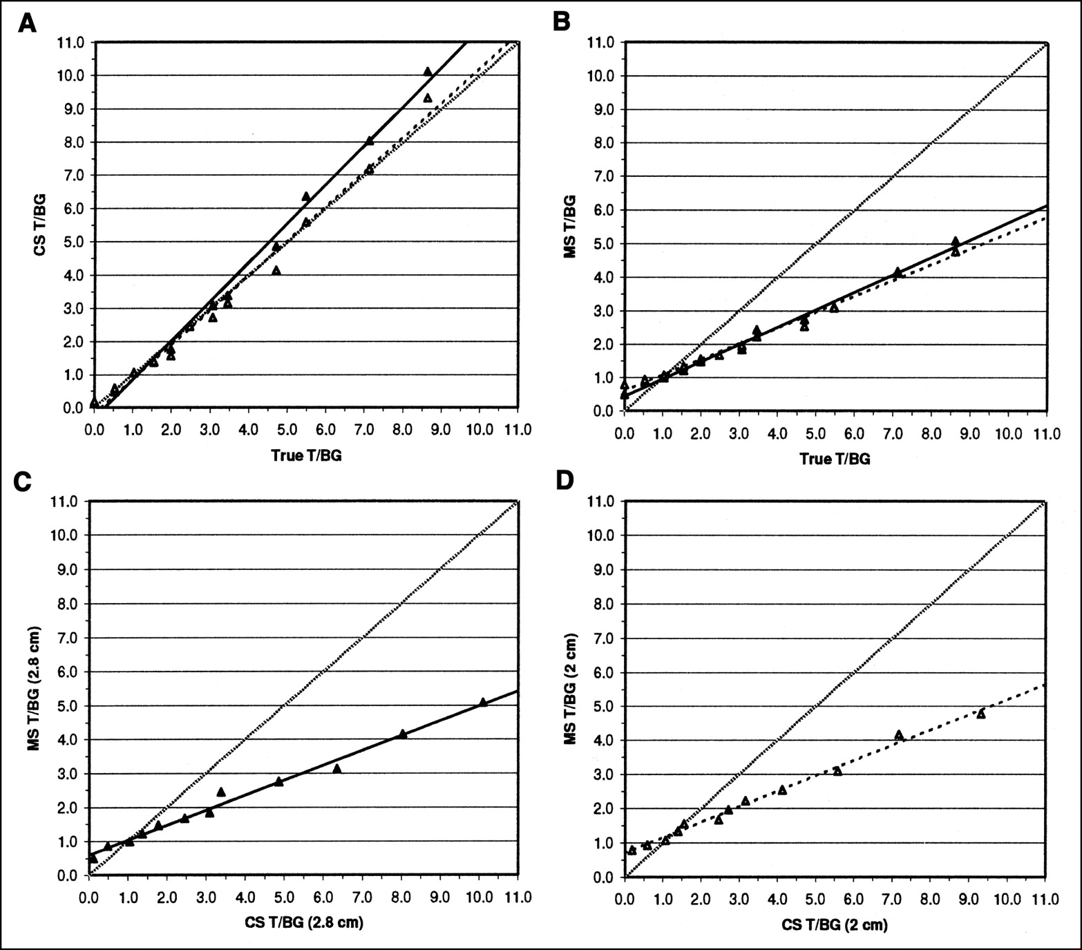
The T/BG ratios recovered by both SPECT systems showed an excellent correlation to the true T/BG given in the phantoms (Fig. 1). As summarized in Table 1, the relationships were sufficiently well fitted by linear regressions, whereas second-order polynomial regressions contributed only slightly to an improved fit in some instances. However, as can be appreciated in Figure 1, the slope of a regression line fitting the data points at the lower end of the examined T/BG range would be less steep, particularly for the CS. We thus performed additional regression analyses for T/BG ratios < 3.5. (This was not pursued for T/BG > 3.5 because of the few data points beyond 3.5.) The results are also summarized in Table 1. Considering all data points, the slopes of the regression lines were significantly less steep for smaller target cylinder diameters for both camera systems (ANCOVA, true T/BG × target cylinder diameter interaction: P < 0.02 for CS; P < 0.05 for MS). (A similar trend was found for the restricted range of T/BG < 3.5: P = 0.085 for CS; P = 0.073 for MS.) The T/BG ratios recovered by both camera systems showed a highly significant linear correlation to each other for both target cylinder diameters (Fig. 1). As summarized in Table 1, a second-order polynomial regression did not contribute or contributed only slightly to an improved fit. Target cylinder diameter did not significantly affect the regression slope (ANCOVA, P > 0.5).

(A and B) Relationship between true T/BG ratios given in phantom studies and T/BG ratios recovered by CS (A) and MS (B). (C and D) Correlation between T/BG ratios recovered in phantom studies by SPECT systems with 2.8-cm target cylinder diameter (C) and SPECT system with 2-cm target cylinder diameter (D). Solid and open triangles represent data points from phantom cylinders of 2.8- and 2-cm diameter, respectively. Solid and dashed lines represent respective linear regression fits. Dotted line represents unity between true and recovered T/BG ratios and both SPECT systems. Table 1 provides regression parameters.
Summary of Regression Analyses in Phantom Studies
Patient Studies
The results of the intra- and interobserver analyses in 123I-FP-CIT patient studies are summarized in Table 2, indicating very high reproducibility and reliability for the used ROI analysis method for all uptake ratios and both SPECT systems. There was no apparent difference between inter- and intraobserver reproducibility. The C/O uptake ratio assessed by the CS tended to show slightly lower reliability, although it was still very high.
Summary of Intra- and Interobserver Analyses
The mean 123I-FP-CIT uptake ratios given by the CS (mean C/O = 5.8 ± 1.4 [coefficient of variation = 24%]; mean P/O = 5.0 ± 1.8 [36%]; mean S/O = 4.8 ± 1.7 [35%]) were significantly higher than the mean uptake ratios given by the MS (mean C/O = 3.4 ± 0.7 [21%]; mean P/O = 3.0 ± 0.8 [27%]; mean S/O = 2.9 ± 0.8 [28%]; all P < 0.0001; no overlap). However, the uptake ratios recovered by both camera systems showed a highly significant linear correlation (Table 3; Fig. 2). For the P/O uptake ratio, the regression slope between both SPECT systems was significantly dependent on the scan order, with a less steep regression slope if the studies were done on the MS first (i.e., group A) (ANCOVA, P < 0.005). The effect of the scan order on the regression slopes for the C/O and S/O uptake ratios failed to reach significance, although showing a tendency toward significance (ANCOVA, P = 0.055 and P = 0.11 for C/O and S/O, respectively). A second-order polynomial regression resulted in only a slightly improved fit (Table 3).
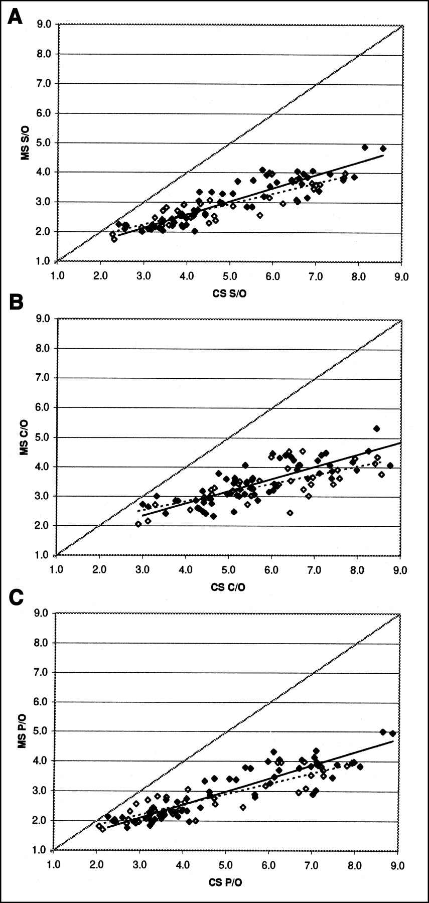
Relationship between S/O (A), C/O (B), and P/O (C) 123I-FP-CIT uptake ratios recovered by CS and MS. Open and solid diamonds represent patients in groups A and B, respectively. Dashed and solid lines represent respective linear regression fits. Dotted line represents unity. Table 3 provides regression parameters.
Summary of Regression Analyses in Patient Studies
DISCUSSION
Several studies have investigated the comparability of different SPECT systems in cerebral blood flow studies. Differences were found not only between single- and multihead cameras (8) but also between different multihead systems (9). Kauppinen et al. (10) showed that single-, dual-, and triple-head systems differed considerably in striatal phantom and 123I-β-CIT studies. None of the systems satisfactorily restored the true binding potential without the use of recovery coefficients. Van Laere et al. (9) performed a data transfer of cerebral blood flow studies from high- to lower-resolution SPECT systems by gaussian filtering of the higher-resolution studies to obtain image characteristics equivalent to the lower-resolution studies. Although such a data transfer is attractive particularly in conjunction with an analysis at a pixel level (e.g., logistic discriminant parametric mapping (11)) for larger centers with high patient throughput, less demanding ROI analyses are better suited for smaller institutions. It has to be questioned whether a transfer of whole datasets is also necessary in SPECT neuroreceptor studies aiming at distinct anatomic regions.
We compared a conventional triple-head SPECT system and a dedicated high-resolution brain SPECT system in phantom and clinical 123I-FP-CIT studies. We found a quasilinear relationship between the T/BG ratios recovered by both cameras and the true T/BG count ratios given in phantoms with targets of the size of the striatum. The smaller regression slope for T/BG ratios at the lower end (T/BG < 3.5) of the data range examined, however, indicates that there is no fully linear relationship. Because a second-order polynomial regression resulted in only marginally improved fits, we appeared justified in primarily using the simpler linear approach. The CS reflected the true T/BG ratios closer than did the MS. The slope of a linear regression between measured T/BG and true T/BG ratios was above unity over the full data range but was slightly below unity for T/BG ratios < 3.5. Thus, the CS system may satisfy the “need for better SPECT imaging” claimed by Kauppinen et al. (10). The respective regression slopes for the MS were considerably below unity for both data ranges. This striking difference between the 2 systems, with the CS performing extraordinarily well, can be attributed to the improved measurement geometry of the CS using an annular sodium iodide single crystal and the dedicated 123I collimator, which provide a considerably higher spatial resolution and sensitivity. Nevertheless, the results of both camera systems showed a close linear relationship that, in contrast to the recovery of the true T/BG ratios, was not significantly affected by the target cylinder diameters examined and was more robust over the full range.
A similar relationship between the 2 SPECT systems was found in vivo using the DAT ligand 123I-FP-CIT. The relationship between the systems was strongest for the P/O uptake ratio (R2 = 0.83 and 0.85 for groups A and B, respectively), whereas it was considerably less strong for the C/O uptake ratio (R2 = 0.52/0.68). The 123I-FP-CIT examinations covered a wide range of uptake ratios (S/O ratio: 1.7–5.0 for MS; 2.0–8.9 for CS), including patients with DAT densities ranging from markedly reduced to normal. Thus, the coefficient of variation was largest for the uptake ratio of the putamen (MS, 27%; CS, 36%) as the most affected uptake ratio in PD, whereas it was lower for the caudate uptake ratio (MS, 21%; CS, 24%). Besides the slightly lower reliability of the C/O uptake ratio assessed by the CS system, this might have favored the detection of a closer relationship between the 2 systems for the P/O uptake ratio. This is an important finding, since the P/O uptake ratio is clinically of the greatest interest—for example, for the early diagnosis of PD. The similar regression parameters of the linear regression analyses between the results of the 2 SPECT systems in 123I-FP-CIT and in phantom studies and the achieved goodness of fit suggest that a linear transformation of striatal 123I-FP-CIT binding measures between the 2 systems may be feasible. However, at this stage, in view of the remaining lack of fit, it is questionable whether the detected relationship is strong enough to allow a data transformation with adequate accuracy in studies investigating subtle variations in healthy volunteers or patient subgroups or in therapy follow-up studies. Although a transformation of reference values may be reasonably accurate for differentiating between subjects with normal and reduced DAT densities (e.g., PD or MSA), in the particular case of 123I-FP-CIT it is questionable whether such a method will be more accurate than the highly accurate visual interpretation (1).
The observation that the linear regression parameters (not the overall goodness of fit) between the 2 SPECT systems were affected by scan order in our patient sample is somewhat intriguing. It is unclear whether this represents a patient sampling effect, although the mean uptake ratios between patient groups A and B were not significantly different (all P > 0.2; Fig. 2), or whether this represents a systematic influence due to the fact that tracer distribution was not sustained at equilibrium conditions during the total scan time. Booij et al. (5) showed that the time to the peak of specific striatal 123I-FP-CIT uptake was less than 3 h in PD patients (0.24–1.91 h) and healthy volunteers (0.76–2.55 h), with patients reaching the peak of specific striatal uptake significantly earlier. There was no significant time trend in specific-to-nonspecific 123I-FP-CIT binding ratios between 3 and 6 h. However, the approximated washout rates tended to be higher for occipital binding (5.6% ± 1.7% and 5.3% ± 2.1% per hour in healthy volunteers and patients, respectively) than for specific striatal binding (3.0% ± 2.0% and 3.7% ± 2.2%, respectively) (5), which would lead to a slight increase of the specific-to-nonspecific uptake ratio with time. This finding could be in line with the effect of scan order on regression slope detected in the present study, that is, slightly less discrepant results for both SPECT systems (regression slope closer to unity) if the SPECT system with lower recovery is used later (midscan-to-midscan interval, approximately 50 min). Besides this possible systematic effect of scan order, the remaining lack of fit in the present study contrasts with the small variability (7.25%) and high interscan correlation (r = 0.98, using a comparable ROI technique) of 123I-FP-CIT uptake ratios found by Booij et al. (12) in a test-retest study. In the view of the high interindividual variability of 123I-FP-CIT tracer kinetics mentioned above (5), the remaining lack of fit might also be at least partially attributed to the different timing of the SPECT studies to be compared. Therefore, the effect of study timing has to be carefully considered. Given the very good test-retest reproducibility of 123I-FP-CIT SPECT studies (12), a 2-d dual-injection protocol seems to be advisable if one seeks to establish highly confident transformation parameters for 123I-FP-CIT studies between certain SPECT systems of interest—for example, of different departments participating in multicenter trials. At this stage, we were interested in whether such a transformation is generally feasible, without aiming at 2 certain SPECT systems. To avoid additional radiation exposure to the patients, we thus used a single-injection protocol.
The effect of study timing will be even more important for tracers with faster kinetics, such as 123I-iodobenzamide, for which a single-injection protocol is not applicable. Our own investigations (single-injection protocol) showed a considerably weaker correlation between the 2 SPECT systems (R2 = 0.20–0.33) in 123I-iodobenzamide studies, most likely because of a more profound effect from different scan times (13).
We intentionally used a somewhat simple analysis method (manually fitted ROI template) that is well suited for clinical routine in every institution and shows high intra- and interobserver reproducibility and reliability. Nevertheless, the correlation between the 2 SPECT systems may be further enhanced by using less observer-dependent methods of data analyses (12,14) or SPECT-MRI coregistration (15). In our experience, the reproducibility of the results of 123I-FP-CIT studies is strongly affected by the placement of the reference ROI. A larger (e.g., whole slice) reference ROI, as used by others (3,16), may be less observer dependent. The analysis may also be more reproducible if one places striatal ROIs in several or all slices showing striatal uptake instead of choosing the slice with the highest striatal uptake (14,16). The slightly lower reliability of the C/O uptake ratios assessed by the CS is most likely due to the small size of the head of the caudate. This structure is more clearly visualized by the CS, so that the C/O uptake ratio is more affected by ROI placement when data acquired with the CS system are analyzed.
An inherent problem with a linear transformation of ligand binding measures between different SPECT systems is their limited resolution. A linear transformation is valid only if the structure of interest is sized similarly to the structures used to define the transformation parameters, since partial-volume effects will lead to a progressive loss of coupling of both SPECT systems if the structure of interest is small in respect to the spatial resolution of the lower-resolution system. This is the case not only in patients with severe structural atrophy (e.g., in Huntington’s disease (4) or MSA) but also when the receptor-bearing “functional” volume is reduced (e.g., in PD with reduced uptake in the putamen). The latter factor might at least in part be responsible for the lower correlation of both SPECT systems with the C/O uptake ratio. However, this principal problem is also encountered when ligand uptake ratios of different individuals studied under identical conditions are compared without applying a partial-volume correction.
CONCLUSION
We found marked differences between 123I-FP-CIT uptake ratios acquired on different modern SPECT systems. The strong correlation between both SPECT systems in phantom studies and (to a lesser extent) in 123I-FP-CIT studies suggests that a linear transformation of striatal uptake ratios between these SPECT systems may be feasible in SPECT receptor studies with ligands such as 123I-FP-CIT. Whether this is achievable with adequate accuracy for clinical purposes needs to be clarified, and if necessary, an optimized analysis method needs to be defined. Proper study timing has to be ensured.
Footnotes
Received Jun. 17, 2002; revision accepted Jan. 20, 2003.
For correspondence or reprints contact: Philipp T. Meyer, MD, Institute of Medicine, Research Center Juelich, 52425 Juelich, Germany.
E-mail: ph.meyer{at}fz-juelich.de
REFERENCES
In this issue


Jump to section
Related Articles
Cited By...
- Evaluation of Collimators in a High-Resolution, Whole-Body SPECT/CT Device with a Dual-Head Cadmium-Zinc-Telluride Detector for 123I-FP-CIT SPECT
- Reproducibility of a Standardized Quantitative Analysis Using Fixed Regions of Interest to Differentiate Movement Disorders on 123I-FP-CIT SPECT
- Clinical Testing of an Optimized Software Solution for an Automated, Observer-Independent Evaluation of Dopamine Transporter SPECT Studies
- 123I-{beta}-CIT SPECT for Imaging Serotonin Transporters in Parkinson's Disease