


Abstract
Pretargeted radioimmunotherapy (RIT) using streptavidin (sAv)-conjugated antibodies before radiolabeled-biotin is a promising approach to improve absorbed dose ratios and achieve high durable remission rates with diminished systemic toxicity. This study compared the immunoscintigraphy, toxicity, and therapeutic efficacy of pretargeted RIT with conventional RIT using an anti-CD20 antibody. Methods: Athymic mice bearing Ramos human Burkitt’s lymphoma xenografts were injected intraperitoneally with a 1F5-sAv conjugate followed 24 h later by a galactosylated, biotinylated clearing agent (CA) and, finally, 3 h later by 111In- or 90Y-labeled 1,4,7,10-tetraazacyclododecane-1,4,7,10-tetraacetic acid (DOTA)-biotin. The comparison groups consisted of mice injected with conventional, directly labeled 111In- or 90Y-1F5. Results: Rapid tumor uptake of radioactivity within 2 h was observed with the pretargeting approach, resulting in high-contrast tumor images at 24 h with minimal blood-pool radioactivity. Although conventional radiolabeled antibodies produced clear tumor images at 24 h, a large amount of radioactivity was present in the blood pool. The tumor-to-blood ratio was 3.5:1 with pretargeting compared with 0.4:1 with conventional 111In-1F5. Pretargeted RIT with 29.6 MBq (800 μCi) 90Y-DOTA-biotin cured 100% of mice with tolerable toxicity, whereas conventional RIT with 90Y-1F5 at a dose of 14.8 MBq (400 μCi) produced no cures, induced profound pancytopenia, and was lethal to all mice. Conclusion: These results suggest that anti-CD20 pretargeted RIT may be superior to conventional radiolabeled antibodies in terms of radioimmunoscintigraphy, toxicity, and therapeutic efficacy for treatment of B-cell lymphomas.
Radioimmunotherapy (RIT) of lymphomas using radiolabeled monoclonal antibodies (mAbs) has produced promising results in multiple clinical trials (1–3). Among the various B-cell surface antigens, CD20, a 35-kDa nonglycosylated phosphoprotein, is the target most commonly used for the RIT of lymphomas. The CD20 antigen is expressed on the surface of almost all mature B-lymphoid cells and on >95% of B-cell lymphomas (4). CD20 is an attractive target for RIT because it is not shed into the circulation, is not rapidly internalized, and is expressed at a high surface density on most lymphomas (5–7). Anti-CD20 antibodies conjugated to 131I or 90Y and administered at nonmyeloablative doses have achieved remissions in 75%–80% of cases, including 35%–40% complete remissions (8–12). High-dose RIT with 131I-anti-B1 (tositumomab) supported by stem cell transplantation has produced objective responses in 85%–90% of cases, including 75%–80% complete remissions (13). Despite its promise, several limitations of RIT have emerged, including the low percentage of injected radiolabeled antibodies that accumulates in tumor sites and the nonspecific irradiation of normal tissues during the long circulation phase of conventional radiolabeled antibodies. Normal tissue toxicities, particularly myelosuppression, have been dose limiting for RIT; therefore, the therapeutic index of systemic RIT might be greatly enhanced if excess nontargeted radiolabeled antibodies could be efficiently cleared from the circulation after maximal accretion of antibody at tumor sites (14). Pretargeted RIT is a promising approach to improve absorbed dose ratios and may correspondingly improve remission rates and remission durations. The pretargeting strategy temporally separates the in vivo tumor localization of the antibody from that of the radionuclide and thereby diminishes systemic toxicities. Pretargeting has been shown to be a more effective approach to obtain selective tumor uptake of radiation with concomitant minimization of radiation doses to normal organs in theoretic analyses, animal models, and clinical trials. Pilot clinical studies have validated the rationale of pretargeting and underscored its promise in relatively radioresistant solid tumor models (15,16); however, only recently have studies investigated this approach in more radiosensitive hematologic malignancies (17–19).
CD20-expressing lymphomas appear to be excellent targets for pretargeted RIT because they are extremely radiosensitive, internalize CD20 antibodies slowly, and have exhibited impressive response rates with chimeric and directly labeled anti-CD20 antibodies (19). Furthermore, temporary eradication of normal B-lymphocytes with malignant lymphoma cells is well tolerated in patients because normal B-lymphocytes killed during therapy are replaced by lymphoid progenitor cells before serious humoral immunodeficiency develops (20).
The suitability of a radionuclide for RIT depends on a variety of factors, such as the nuclides’s radiophysical properties, the target tumor’s morphology and physiology, the antibody’s targeting kinetics, the in vivo stability of the nuclide, the nuclide’s accessibility, and, finally, the availability of simple and efficient clinical-scale radiolabeling methods. 131I and 90Y are β-particle–emitting radioisotopes that have been most widely used for RIT trials. 131I has been used in the earliest clinical RIT trials because of its ready availability, low cost, simple protein labeling chemistry, the presence of γ-emissions suitable for gamma-camera imaging, and an 8-d half-life (21). 90Y is a pure β-emitter and emits high-energy, long-range β-particles that make 90Y suitable for the irradiation of large tumor masses. Moreover, 90Y and similar metallic radionuclides are “residualizing” labels and remain securely sequestered inside lysosomes after endocytosis (21,22).
Various systems have been proposed for the pretargeted delivery of radioactivity to tumors (23). Biotinylated or avidin-conjugated mAbs that bind radiolabeled avidin or biotin, respectively, have been found to be effective methods for the pretargeted delivery of radioactivity to tumors. The avidin-biotin system has many advantages, including the very high affinity of avidin and streptavidin (sAv) for biotin and the multivalency of the system (each sAv or avidin can bind 4 biotin molecules) (19,24). Our laboratory is currently investigating a 2-step pretargeting approach using sAv-conjugated anti-CD20 mAbs in an athymic mouse xenograft system. This report documents the superior tumor localization, imaging, toxicity, and therapeutic efficacy of a pretargeted anti-CD20 antibody compared with the same antibody administered after conventional, direct radiolabeling.
MATERIALS AND METHODS
Cell Lines and Lymphoma Xenografts
The human Ramos B-lymphoma cell line (American Type Culture Collection, Bethesda, MD) was maintained in logarithmic-phase growth in RPMI 1640 medium containing 10% heat-inactivated fetal calf serum in a 5% CO2 incubator. Cell viability was determined by trypan blue exclusion. Cell viabilities of >90% were required for injection into mice.
Six- to 8-wk-old female BALB/c nude mice (B & K Universal, Kent, WA) were injected subcutaneously with 10 × 106 Ramos cells in each flank. After 7–10 d, palpable tumors appeared and mice with similar tumor sizes (∼5-mm diameter) were used for experiments. Tumor-bearing mice were placed on a biotin-free diet (Harlan Teklad, Madison, WI) for 4–7 d before injection of mAb-sAv conjugates and radiobiotin. Animal studies were conducted under the supervision of veterinarians from the University of Washington Comparative Medicine Department or the Animal Health Resources Department of the Fred Hutchinson Cancer Research Center.
Antibodies and sAv Conjugates
The murine anti-human CD20 IgG2a mAb 1F5 was produced in a hollow fiber bioreactor system in the Fred Hutchinson Cancer Research Center Monoclonal Antibody Facility (Seattle, WA). The 1F5 hybridoma was a gift of Dr. Clay Siegall (Seattle Genetics Inc., Seattle, WA). Two irrelevant antibodies, isotype-matched NR-LU-10 (gift from NeoRx Corp., Seattle, WA) and G3G6 (gift from Dr. Dana Matthews, Fred Hutchinson Cancer Research Center, Seattle, WA), were used as nonspecific controls. A conjugate of recombinant sAv and 1F5 was prepared as described (19) using a variation of the method of Hylarides et al. (25). Briefly, sAv was concentrated to 20 mg/mL and reacted with a 3 molar excess of succinimidyl 4-[N-maleimidomethyl]cyclohexane-1-carboxylate (SMCC; Pierce, Rockford, IL) for 30 min at pH 8.0. Unreacted SMCC and reaction by-products were removed using G-25 chromatography equilibrated in phosphate-buffered saline (PBS). The 1F5 was concentrated to 10 mg/mL at pH 8.0 and dithiothreitol (Pierce) added to a final concentration of 20 mmol/L. After a 30-min reaction, residual dithiothreitol was removed from the reduced 1F5 using G-25 chromatography as described. After G-25 purification, sAv contained 1.5 reactive maleimides and 1F5 contained 8.4 thiols per molecule as determined using the 5,5′-dithiobis-(2-nitrobenzoic acid) (Pierce) assay described by Hylarides et al. (25).
Sodium chloride was added to the reaction mixture to a final concentration of 0.4 mol/L, glycine was added to 0.05 mol/L, and the pH was adjusted to 9.2 with 1N NaOH. The crude conjugation mixture was applied over an iminobiotin column (Pierce) equilibrated in 0.05 mol/L glycine, 0.5 mol/L NaCl, pH 9.2. Unreacted 1F5 flowed through the column and unreacted sAv and 1F5-sAv conjugate eluted with 0.2 mol/L acetate buffer, pH 4.0. The eluted fraction was diluted by buffer exchange to a conductivity of <2.5 mS/cm with distilled H2O and the pH was adjusted to 6.5 with 1N NaOH. The conjugation mixture obtained from the iminobiotin column was purified by cation-exchange chromatography using a Fractogel EMD SO3− (S) column (EM Separations Technology, Gibbstowin, NJ) equilibrated in 20 mmol/L sodium phosphate buffer, pH 6.5, as described (19). After the load, the column was washed with equilibration buffer to remove the unreacted sAv. The 1F5-sAv conjugate was eluted with 90 mmol/L NaCl in equilibration buffer. The maximal yield of 1F5-sAv after dual-column purification was 35% (mg protein product/mg protein starting materials). The final 1F5-sAv conjugate contained 80%–85% 1:1 1F5-sAv conjugates, 5%–10% 1:2 1F5-sAv conjugates, and 6%–10% molecules with higher ratios of sAv:1F5. Aggregated conjugate eluted at higher concentrations of NaCl. The immunoreactivity of the purified conjugate was determined using a competitive enzyme-linked immunosorbent assy. The biotin binding capacity was determined by displacement of 2-(4′-hydroxyphenylazo)benzoic acid (Aldrich, Milwaukee, WI) from sAv as described (26). The biotin binding capacity of 1F5-sAv was 5 mol of biotin/mol of conjugate. These data confirm high-performance liquid chromatography (HPLC) results showing that some 1F5-sAv conjugates contain 2 or more sAv moieties per antibody molecule.
Clearing Agent
A completely synthetic clearing agent (CA) was used to remove excess unbound mAb-sAv conjugates from the bloodstream before radio-biotin administration. The synthesis and characterization of this reagent have been published elsewhere (27,28). This agent, designated biotin-LC-NM-(Gal-NAc)16 (molecular weight = 8651.6), is a bifunctional moiety consisting of biotin joined, through a modified aminocaproyl spacer, to the core of a 4-generation dendrimeric backbone formed using repetitive bifunctional units (27,28). The outer dendrimeric shell is functionalized with 16 modified N-acetyl-galactosamine residues through aminopentyl linkages. The agent is administered in stoichiometric dose excess to the mAb-sAv conjugate. It has a volume of distribution that allows it to access and bind to vascular and extravascular conjugates. The N-acetyl-galactosamine residues have a high affinity for hepatic asialoglycoprotein receptors, which mediate the rapid hepatic clearance of residual 1F5-sAv conjugates from the bloodstream and their endocytosis into liver cells. Through competitive in vitro binding assays, it has been determined that, although the affinity of this CA for sAv is quite high, it is nonetheless significantly lower than that of natural biotin or of the subsequently administered radionuclide-biotin-chelate ligand (27,28). Thus, although the CA may also bind significantly to extravascular conjugate under conditions of high doses, it does not compromise the binding of subsequently administered biotin-chelate ligand. Numerous studies have been performed documenting that the biotin-chelate ligand can effectively compete with the CA under in vivo conditions and time frames (27,28).
Radiolabeling of mAbs
Radioiodination of mAb-sAv with 131I or 125I (NEN Life Science Products, Inc., Boston, MA) was performed using the chloramine-T method as described (29).
mAbs were radiolabeled with 111In or 90Y (NEN Life Science Products, Inc., Boston, MA) using p-isothiocyanatobenzyl-1,4,7,10-tetraazacyclododecane-1,4,7,10-tetraacetic acid (p-SCN-Bz-DOTA; Macrocyclics, Richardson, TX) by the method of Mirzadeh et al. (30). Buffer solutions were prepared using metal-free reagents, and storage vials and the mAbs were made metal free using Chelex 100 resin (Bio-Rad Laboratories, Hercules, CA). The radiochemical purity of the conjugates was consistently >95% by thin-layer chromatography.
111In- or 90Y-DOTA-Biotin
The bifunctional ligand DOTA-biotin, a gift from NeoRx Corp. (Seattle, WA), was synthesized as described (31). Carrier-free 111In-chloride (NEN Life Science Products), 0.02–0.5 mL in 0.04 mol/L HCl, was diluted with 0.5 mL of 2 mol/L ammonium acetate, pH 5. DOTA-biotin, 0.1–1 mg, was added, and the solution was heated for 30 min at 80°C. Diethylenetriaminepentaacetic acid (DTPA), 0.05 mL of a 0.1 mol/L solution, was added to chelate any unbound 111In. Carrier-free 90Y-Cl3 (NEN Life Science Products), 0.02–0.2 mL in 0.05 mol/L HCl, was diluted with 2 mol/L ammonium acetate, pH 5, to a total volume of 0.4 mL. Ascorbic acid, 0.05 mL of a 0.5 g/mL solution, and 0.1 mL of 10 mg/mL DOTA-biotin were added, and the solution was heated at 80°C for 1 h. DTPA, 0.05 mL of a 0.1 mol/L solution, was added to chelate any unbound radiometal. The labeling efficiencies for 111In- and 90Y-DOTA-biotin were >94% as assessed using an avidin-bead method and HPLC (19).
Radioimmunoscintigraphy
Nude mice bearing lymphoma xenografts were divided into 2 groups. The pretargeted group was injected intraperitoneally with 1.4 nmol of 1F5-sAv (300 μg) followed 24 h later with 5.8 nmol of CA. Three hours later, the mice were anesthetized with intraperitoneal pentobarbital (50 mg/kg of body weight) and were injected with 1.2 nmol (1 μg) of DOTA-biotin labeled with 11.1 MBq (300 μCi) of 111In in a tail vein and gamma-camera imaging was performed after 0, 2, and 24 h. The control or conventional radioimmunoscintigraphy group of mice were treated by injecting 1.4 nmol (215 μg) of 1F5 directly labeled with 11.1 MBq (300 μCi) of 111In. After the injection of 111In-biotin in pretargeted mice and 111In-mAb in conventional radioimmunoscintigraphy mice, the mice were imaged with a model 400AT gamma camera (General Electric Medical Systems, Milwaukee, WI) equipped with a high-energy collimator at 0-, 2-, and 24-h time points. After 24 h, mice were euthanized, and tumors and normal tissues were excised and assayed for radioactivity.
RIT
To compare the therapeutic efficacies of pretargeted and conventional radiolabeled antibodies, groups of 5 tumor-bearing mice were injected with 1.4 nmol (215 μg) and 7.4 or 14.8 MBq (200 or 400 μCi) of directly labeled 90Y-DOTA-1F5, equimolar amounts (300 μg) of 1F5-sAv or NR-LU-10-sAv conjugates, followed 24 h later by 5.8 nmol (50 μg) of CA and 3 h later by 1.2 nmol (1 μg) of DOTA-biotin labeled with 14.8 or 29.6 MBq (400 or 800 μCi) of 90Y. Mice were monitored every other day for general appearance, tumor volume, and body weight. Mice were euthanized if tumors became large enough (1–1.5 cm in diameter) to cause obvious discomfort or impair ambulation. Differences in mean tumor sizes were analyzed with the Student t test and differences were considered significant at the P < 0.05 level.
Toxicity Analysis
Hematopoietic, renal, and liver toxicity were assessed in BALB/c athymic mice. To assess toxicities beyond 10 d without the confounding effects of euthanasia for rapidly growing tumors, it was necessary to perform these studies in mice that were not bearing tumors. Mice were injected with 1.4 nmol (215 μg) and 7.4 or 14.8 MBq (200 or 400 μCi) of directly labeled 90Y-DOTA-1F5, equimolar amounts (300 μg) of 1F5-sAv conjugates, followed 24 h later by 5.8 nmol (50 μg) of CA and 3 h later by 1.2 nmol (1 μg) of DOTA-biotin labeled with 14.8 or 29.6 MBq (400 or 800 μCi) of 90Y. Blood samples were drawn on days 0, 7, 14, 28, and 35, and the number of leukocytes, neutrophils, and platelets was measured by a CELL-DYN 3500R instrument (model 3500 CS; Abbott Laboratories, Inc., North Chicago, IL). To evaluate hepatotoxicity and nephrotoxicity, serum aspartate aminotransferase (AST), alanine aminotransferase (ALT), and creatinine (CRE) levels were measured on days 0, 7, 14, and 28 using a Synchron LX 20 instrument (Beckman Coulter, Inc., Fullerton, CA). Duplicate samples were collected from each mouse and the average was reported.
RESULTS
Effects of Synthetic CA on Circulating 1F5-sAv Conjugate
The blood clearance of 1.4 nmol of 125I-1F5-sAv was assessed in BALB/c athymic mice injected with a synthetic biotinylated N-acetyl-galactosamine–containing CA that was administered 24 h after the anti-CD20 immunoconjugate (Fig. 1). The blood concentration of 125I-1F5-sAv dropped from 20.44 %ID/g (percentage injected dose per gram) to 1.67 %ID/g after 5.8 nmol of CA and to 2.13 %ID/g after 11.6 nmol of CA. The CA effectively reduced the blood concentration by 91% within 30 min of administration of 5.8 nmol of CA and by 89% after 11.6 nmol of CA. The blood clearance of 1F5-sAv was also demonstrated visually using gamma-camera imaging (model 400AT; General Electric) of mice injected with 5.8 nmol of CA 24 h after the injection of 131I-1F5-sAv, showing hepatic clearance of the 1F5-sAv-CA complex (Fig. 2).

Effects of CA on circulating 1F5-sAv conjugate. BALB/c mice (n = 4) were injected with 1.4 nmol of 125I-1F5-sAv conjugate at time 0 and 24 h later with saline (•) or 5.8 nmol of CA (□) or 11.6 nmol of CA (▴). Blood samples were collected at 6, 22, 25, 27, 29, and 44 h after 125I-1F5-sAv injection and assayed in gamma counter. Data represent mean percentage injected dose per gram (%ID/g) ± SD.

Gamma-camera images of mice injected with 1.4 nmol of 131I-1F5-sAv at 24 h (just before CA injection) (A), 25 h (1 h after CA injection) (B), and 48 h (24 h after CA injection) (C). Images are shown at same camera intensity settings. CA was injected 24 h after 131I-1F5-sAv administration. Arrows indicate radioactivity in thyroid (Th), blood pool (B), and liver (L).
Radioimmunoscintigraphy
Gamma-camera imaging was performed in athymic mice bearing Ramos lymphoma xenografts 2 and 24 h after injection of 111In-DOTA-biotin (Fig. 3). Rapid uptake of radioactivity was observed in tumors after 2 h in the pretargeted group (Fig. 3B) as well as rapid urinary excretion of unbound radiobiotin, manifested by a large amount of radioactivity in the bladder. In the conventional RIT group, most of the radioactivity at 2 h was found in the blood pool and no tumor localization was observed (Fig. 3A). By 24 h, most of the bladder radioactivity had cleared in the pretargeted group, resulting in high-contrast tumor images (Fig. 3D). Although tumors were visible in the conventional RIT group at 24 h, a large amount of radioactivity was also present in the blood pool (Fig. 3C). After the 24-h scan, mice were euthanized and the distribution of radioactivity in tumors and normal organs was assayed. The pretargeting approach resulted in superior biodistributions of radioactivity compared with those in the conventional RIT group, as shown in Figure 4A. With pretargeting, the tumor content of 111In-DOTA-biotin was 4.35 ± 0.30 %ID/g at 24 h, whereas the blood content was only 1.26 ± 0.59 %ID/g. In contrast, the tumor uptake of conventional 111In-1F5 was 4.64 ± 0.67 %ID/g at 24 h with a blood content of 10.36 ± 4.73 %ID/g. Tumor-to-blood ratios were 3.5:1 at 24 h with pretargeting compared with 0.4:1 at 24 h with conventional targeting (Fig. 4B). These results are concordant with our earlier comparative biodistribution experiments (19).

Gamma-camera images of Ramos xenograft-bearing athymic mice injected with either directly labeled 111In-1F5 antibody (A and C) or pretargeted with 1F5-sAv conjugate followed 24 h later by CA and then by 111In-DOTA-biotin (B and D). Images are shown at same camera intensity settings. Images of mice are shown 2 h after injection of radioactivity (A and B) and 24 h after injection of radioactivity (C and D). Arrows indicate radioactivity in tumor (T), blood pool (B), and urinary bladder (U).
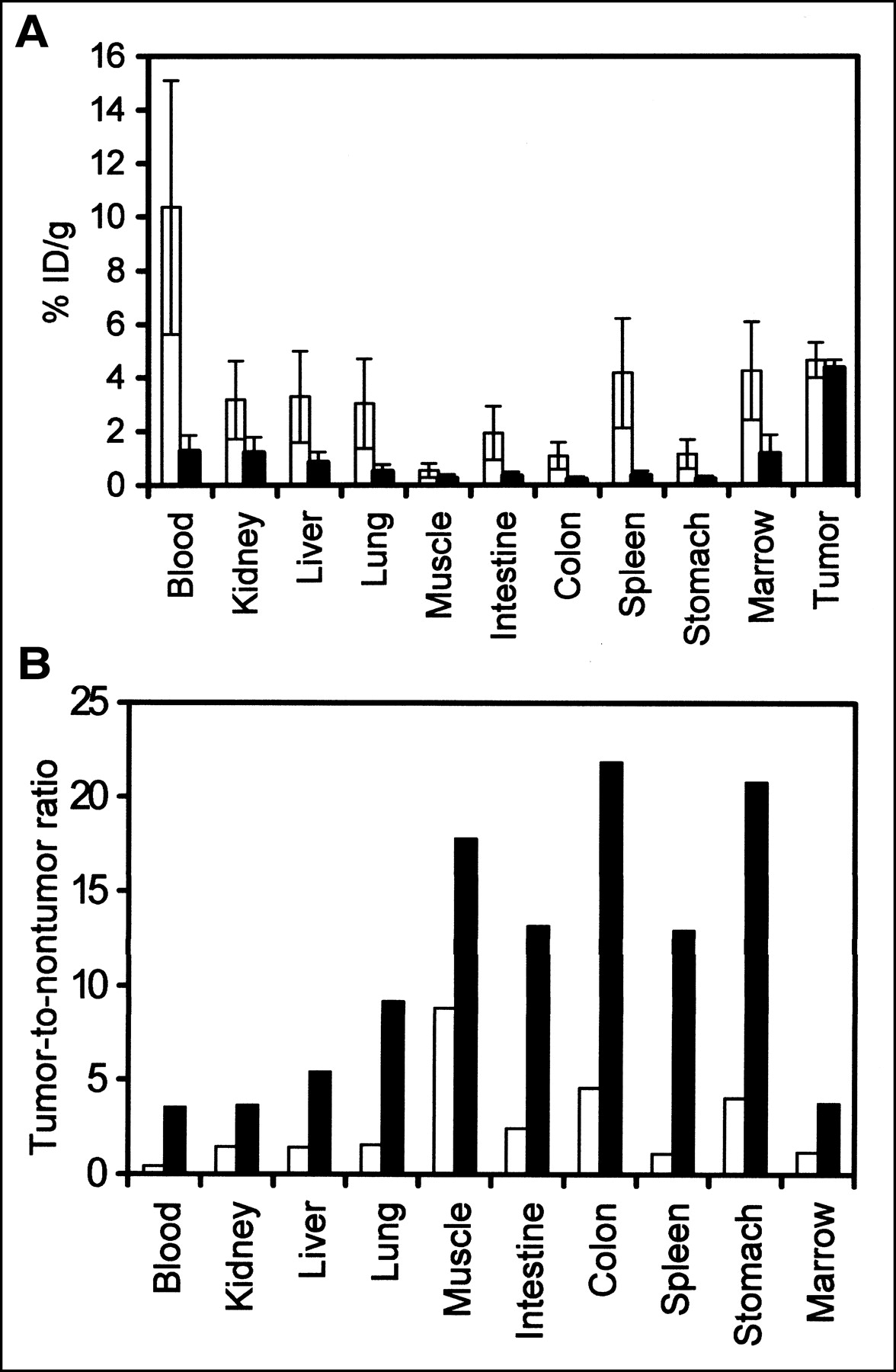
Biodistributions of radioactivity (A) and tumor-to-normal organ ratios (B) of Ramos xenograft-bearing mice injected either with directly labeled 111In-1F5 (□) or with 1F5-sAv followed 24 h later by CA and then 1 h later with 111In-DOTA-biotin (▪). Data in A were obtained 24 h after injection of radioactivity and are shown as %ID/g ± SD. Data represent the mean of 3 mice in each group.
RIT
On the basis of the promising findings of the imaging and biodistribution studies, we performed RIT experiments to evaluate whether the pretargeting method would provide enhanced therapeutic efficacy compared with conventional 1-step RIT. Groups of 5 lymphoma-bearing mice were injected with 1.4 nmol of 1F5-sAv followed 24 h later by 5.8 nmol of CA and 3 h later by 1.2 nmol of 90Y-DOTA-biotin labeled with 14.8 or 29.6 MBq (400 or 800 μCi) per mouse. Mice injected with 1.4 nmol and 7.4 or 14.8 MBq (200 or 400 μCi) of directly labeled 90Y-DOTA-1F5 per mouse were used as a comparison. The control groups included mice injected with 1.4 nmol of the nonspecific NR-LU-10-sAv conjugate + 5.8 nmol of CA + 29.6 MBq (800 μCi) of 90Y-DOTA-biotin and mice injected only with PBS.
Mice treated with PBS alone or with the control NR-LU-10-sAv conjugate followed by 29.6 MBq (800 μCi) of 90Y-DOTA-biotin experienced no tumor regression and were euthanized between days 13 and 16 because of the exponential growth of their tumor xenografts (Fig. 5A). Mice treated with 7.4 MBq (200 μCi) of conventional 90Y-DOTA-1F5 exhibited a tumor growth delay of ∼29 d but no complete remissions were observed and all mice eventually required euthanasia as a result of progressive tumor growth (Fig. 5A). Mice treated with 14.8 MBq (400 μCi) of conventional 90Y-DOTA-1F5 experienced a very slow tumor growth but all mice in this group experienced an average 17.3% ± 6.32% loss of their initial body weight and died by day 13 because of toxicity (Fig. 5B).
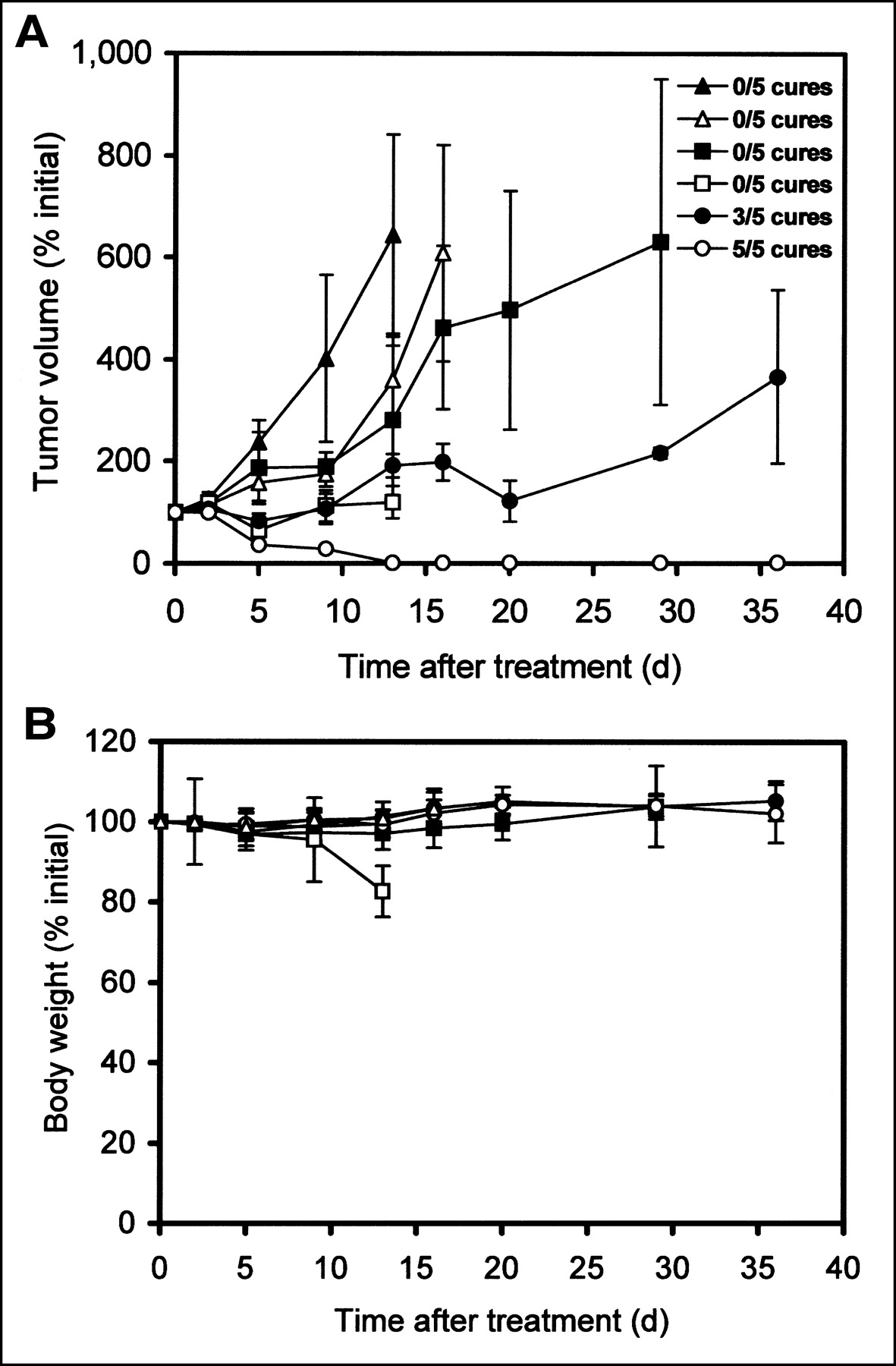
Regression of lymphoma xenografts (A) and loss of body weight (B) after conventional or pretargeted RIT. BALB/c nude mice bearing Ramos lymphoma xenografts were injected with saline alone (▴), with control NR-LU-10-sAv followed by CA and later by 29.6 MBq (800 μCi) of 90Y-DOTA-biotin (▵), with 7.4 MBq (200 μCi) (▪) or 14.8 MBq (400 μCi) (□) of directly labeled 90Y-1F5, or with 1F5-sAv followed by CA and later by 14.8 MBq (400 μCi) (•) or 29.6 MBq (800 μCi) (○) of 90Y-DOTA-biotin. Data represent mean percentage ± SD of initial values (n = 5).
In contrast, mice in the pretargeted groups achieved better tumor responses with less toxicity. Three of 5 mice treated with pretargeted 1F5-sAv + 14.8 MBq (400 μCi) of 90Y-DOTA-biotin obtained complete tumor remissions, but the other 2 mice did not respond (Fig. 5A). In the pretargeted group receiving 29.6 MBq (800 μCi) 90Y-DOTA-biotin, all 5 mice achieved complete tumor remissions (P < 0.05) by day 13 (Fig. 5A) without any regrowth of the tumors for >125 d. No significant weight loss or visible signs of toxicity were observed in these mice treated with pretargeted 1F5-sAv + 14.8 or 29.6 MBq (400 or 800 μCi) of 90Y-DOTA-biotin (Fig. 5B). The differences in tumor growth observed between the pretargeting groups and the conventional RIT groups were statistically significant (P = 0.008 for pretargeting with 400 μCi vs. conventional RIT with 200 μCi at 20 d; P = 0.0003 for pretargeting with 800 μCi vs. conventional RIT with 400 μCi at 9 d; and P = 0.004 for pretargeting with 400 μCi vs. conventional RIT with 400 μCi at 13 d).
Toxicity Analysis
Hematopoietic, hepatic, and renal toxicities were assessed in BALB/c, athymic mice injected with either conventional or pretargeted RIT protocols. To assess toxicities beyond 10 d without the confounding effects of euthanasia for rapidly growing tumors, it was necessary to perform these studies in mice that did not bear tumors. As shown in Figure 6A, mice injected with 7.4 MBq (200 μCi) of conventional 90Y-DOTA-1F5 exhibited a significant decrease (79.2% ± 0.73%) in the neutrophil count by day 7, which persisted for a week, but the neutrophil count subsequently rebounded to the initial normal value by day 28. Mice injected with 14.8 MBq (400 μCi) of conventional 90Y-DOTA-1F5 exhibited an even more profound decrement in the neutrophil count (87.7% ± 0.69%) by day 7, and eventually all mice in this group died between days 10 and 12 of presumed infections. Neutropenia evolved more slowly and was less marked in the pretargeted groups injected with either 14.8 or 29.6 MBq (400 or 800 μCi) of 90Y-DOTA-biotin (Fig. 6A). The neutrophil nadir occurred on day 14 in both pretargeted groups, with a mean nadir neutrophil count of 0.63 ± 0.32 × 10³/mm3 in the 14.8-MBq (400 μCi) group and 0.31 ± 0.27 × 10³/mm3 in the 29.6-MBq (800 μCi) group. In the pretargeted group injected with 29.6 MBq (800 μCi) of 90Y-DOTA-biotin, a mean decrease of 85.3% ± 0.53% in the neutrophil count was observed, similar to the decrement observed with 7.4 MBq (200 μCi) of conventional 90Y-DOTA-1F5. In both pretargeted groups, the original normal neutrophil counts were regained by day 35. A similar pattern was observed with the total leukocyte counts (Fig. 6B) and platelet counts (Fig. 6C). Thrombocytopenia was significantly less marked with 29.6 MBq (800 μCi) of pretargeted 90Y-DOTA-biotin than with 7.4 MBq (200 μCi) of conventionally targeted 90Y-1F5 (7.79 ± 0.66 × 10⁵/mm3). No significant changes in the hematocrit (Fig. 6D) were observed in either the 14.8- or 29.6-MBq (400 or 800 μCi) pretargeted group. However, in the conventional RIT group given 7.4 MBq (200 μCi) of 90Y-DOTA-1F5, a marked reduction in the hematocrit was observed with a nadir hematocrit of 25.4% ± 1.8% (46.4% decrease). Anemia resolved by day 35. The hematocrit fell significantly (15.2%) by day 7 in the 14.8-MBq (400 μCi) conventional RIT group, but complete assessment was not possible because all mice died of hematopoietic toxicity by day 12.
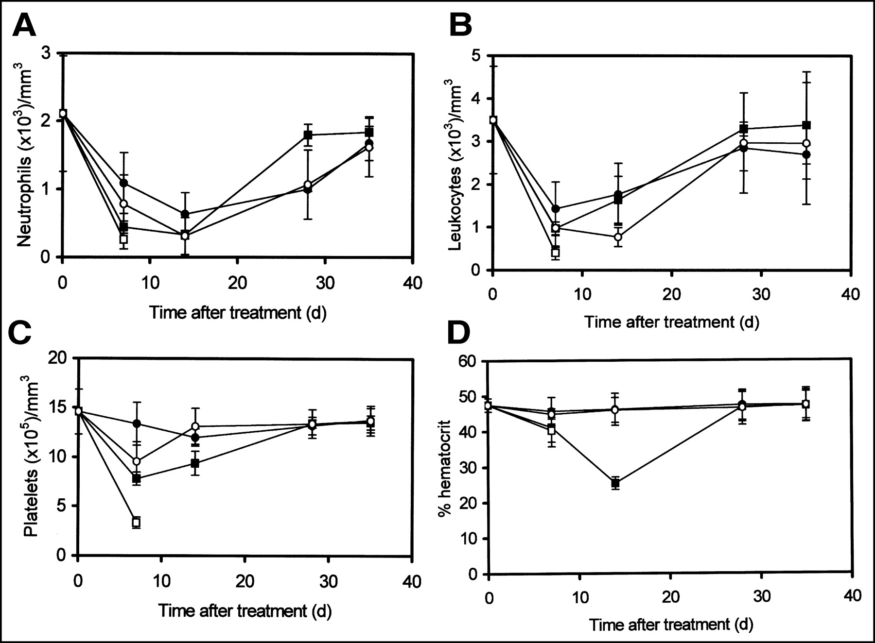
Hematologic toxicity of mice as indicated by neutrophil counts (A), leukocyte counts (B), platelet counts (C), and hematocrit (D). BALB/c mice were injected with 7.4 MBq (200 μCi) (▪) or 14.8 MBq (400 μCi) (□) of directly labeled 90Y-1F5 or with 1F5-sAv followed by CA and later by 14.8 MBq (400 μCi) (•) or 29.6 MBq (800 μCi) (○) of 90Y-DOTA-biotin. Data represent mean ± SD of 4 mice in each group.
To evaluate hepatotoxicity and nephrotoxicity, AST, ALT, and CRE levels were measured on days 0, 7, 14, and 28. No major changes in the levels of AST, ALT, and CRE were observed in any group during the 28 d of observation (Table 1).
Hepatotoxicity and Nephrotoxicity Analysis of Mice Injected with Directly Labeled 90Y-1F5 and Pretargeted 1F5-sAv + CA + 90Y-DOTA-Biotin
DISCUSSION
This report demonstrates that pretargeted anti-CD20 RIT provides efficient tumor localization with improved tumor-to-normal tissue ratios, generates higher quality tumor imaging, and induces higher durable remission rates and reduced toxicities compared with conventional RIT. The rapid clearance of unbound circulating anti-CD20-sAv immunoconjugate from the bloodstream by the CA and the rapid renal elimination of excess 111In-DOTA-biotin in the pretargeted RIT approach contributed to the superiority of the tumor images with the pretargeting method. Scintigraphic images showed a rapid tumor uptake of 111In-DOTA-biotin in the pretargeted group within 2 h of 111In-DOTA-biotin injection, whereas most of the nontargeted radioactivity was rapidly eliminated in the urine, resulting in highly specific tumor images at 24 h. In contrast, tumor images generated using conventional RIT with 111In-1F5 were confounded by high residual levels of radioactivity persisting in the blood and other nontarget organs.
The markedly superior biodistributions of radioactivity using the anti-CD20 pretargeting approach demonstrated by radioimmunoscintigraphy and by γ-counting of excised organs suggest that pretargeting would also afford superior therapeutic efficacy and diminished toxicity compared with conventional RIT. These predictions have been confirmed by the RIT experiments presented in this article and in previous studies (17,19). Conventional 90Y-1F5 antibodies were unable to cure animals bearing Ramos lymphoma xenografts at sublethal radiation doses of <14.8 MBq (<400 μCi). In marked contrast, tumor-bearing mice were reliably cured using pretargeted anti-CD20 RIT at optimal doses. Importantly, the curative doses of 1F5-sAv + 90Y-DOTA-biotin used were well tolerated by mice.
Various RIT studies have clearly shown that directly labeled mAbs generally induce marked levels of hematotoxicity because of the presence of high levels of radioactivity in the bloodstream for prolonged periods of time (32,33). Our study demonstrates that this hematotoxicity can be reduced significantly using pretargeting, which is capable of delivering higher amounts of radioactivity to tumors without increasing marrow exposure to radioactivity. In this study, hematologic toxicity was significantly lower with pretargeted RIT, even after the administration of 29.6 MBq (800 μCi) of 90Y, which was at least twice a 100% lethal dose (14.8 MBq or 400 μCi of 90Y) of the directly conjugated antibody. In contrast, the prolonged serum half-life of directly labeled 1F5 led to substantial toxicity at 7.4 MBq (200 μCi) and fatal toxicities at 14.8 MBq (400 μCi) of 90Y. Detailed serial analyses of hematologic parameters confirmed that leukopenia, neutropenia, anemia, and thrombocytopenia were all significantly reduced in mice treated with the pretargeting approach compared with conventional RIT. The reduced toxicity of pretargeted RIT enabled the safe delivery of radioactivity at twice the lethal dose of directly labeled antibodies and resulted in ∼90% apparent cures. The CA appears to play a key role in the removal of excess 1F5-sAv immunoconjugate from circulation and, thereby, improves the tumor-to-normal organ ratios, leading to the enhancement of the therapeutic index.
Significant nephrotoxicity and hepatotoxicity were not observed in these experiments in either the pretargeted or the conventional RIT group. However, delayed hepatotoxicity, nephrotoxicity, and radiation cystitis might occur with longer follow-up as a result of the urinary excretion of large doses of 90Y-DOTA-biotin. To assess this possibility we are conducting long-term experiments with mice treated with pretargeted RIT and large doses of 90Y-DOTA-biotin up to 44.4 MBq (1,200 μCi) with serial measurements of liver transaminases and serum CRE over a period of 1 y. At the end of the year, mice will be euthanized and the liver, bladder, and kidneys will be examined histologically for evidence of radiation injury or fibrosis.
The promising results of our experiments using murine xenograft models suggest that clinical trials using pretargeted RIT might permit substantial dose escalation of CD20-directed RIT to radiation doses that would produce higher rates of complete remission and improved cure rates in lymphoma patients with tolerable toxicity and without the need for stem cell rescue. Despite the fact that pretargeting appears very promising, this method also has limitations. Among the disadvantages of pretargeting are the requirement for separate, sequential injections at defined time intervals and the immunogenicity of sAv (34). Other reservations can also be identified that mandate caution in extrapolating these murine experiments to humans. First, radioimmunoconjugates may have more uniform tumor uptake in this mouse xenograft system than that in humans because of the improved vascularity of the tumor xenografts. Second, mouse B-lymphocytes do not bind anti-CD20 antibodies but human B-lymphocytes will, which might adversely impact targeting in patients unless circulating B-cells are precleared by injection of nonradioactive anti-CD20 antibody before RIT. Third, the relatively long pathlength of β-emissions of 90Y in relation to the size of the mouse and the size of the tumor xenografts may be considered disadvantageous. On the other hand, if one considers the enhanced toxicity to a greater volume of normal tissue in the mouse model from β-particles emanating from the tumors that deposit their energy outside the xenograft, mouse xenograft studies using 90Y-immunoconjugates can be conservative predictors of human results (35). Despite these caveats, we believe that this investigation, and those of other investigators (17,19,35), has firmly established that pretargeted RIT can provide remarkable discrimination between tumor and normal tissue and that this enhanced targeting translates to superior RIT efficacy.
CONCLUSION
The results of our study demonstrate the superiority of pretargeted RIT compared with conventional RIT because of the higher uptake of radioactivity; more rapid elimination of radioactivity from the circulation and whole body, resulting in improved tumor-to-normal organ ratios of absorbed radioactivity; the ability to deliver higher doses of radioactivity, with tolerable toxicity to marrow and normal organs; improved complete remission rates; and enhanced survival of tumor-bearing mice. The superiority of pretargeted RIT over conventional RIT merits further preclinical and clinical studies, particularly with novel pretargeting reagents such as engineered anti-CD20-sAv fusion proteins (17), single-chain antibodies from phage display libraries (36), or bispecific antibodies recognizing bivalent haptens (37,38).
Acknowledgments
This study was supported by National Institutes of Health grant R01 CA76287 and by gifts from the Dr. Penny E. Peterson Endowment and the Hext Family Foundation.
Footnotes
Received Mar. 29, 2002; revision accepted Sep. 25, 2002.
For correspondence or reprints contact: Oliver W. Press, MD, PhD, Fred Hutchinson Cancer Research Center, D3-190, 1100 Fairview Ave. N., Seattle, WA 98109.
E-mail: press{at}u.washington.edu
REFERENCES
In this issue


Jump to section
Related Articles
Cited By...
- A Self-Assembling and Disassembling (SADA) Bispecific Antibody (BsAb) Platform for Curative Two-step Pretargeted Radioimmunotherapy
- Therapy of Myeloid Leukemia using Novel Bispecific Fusion Proteins Targeting CD45 and 90Y-DOTA
- Venetoclax Synergizes with Radiotherapy for Treatment of B-cell Lymphomas
- Comparative Analysis of Bispecific Antibody and Streptavidin-Targeted Radioimmunotherapy for B-cell Cancers
- A Preclinical Model of CD38-Pretargeted Radioimmunotherapy for Plasma Cell Malignancies
- Conventional and pretargeted radioimmunotherapy using bismuth-213 to target and treat non-Hodgkin lymphomas expressing CD20: a preclinical model toward optimal consolidation therapy to eradicate minimal residual disease
- Pretargeting CD45 enhances the selective delivery of radiation to hematolymphoid tissues in nonhuman primates
- A comparative analysis of conventional and pretargeted radioimmunotherapy of B-cell lymphomas by targeting CD20, CD22, and HLA-DR singly and in combinations
- Improved Therapeutic Results by Pretargeted Radioimmunotherapy of Non-Hodgkin's Lymphoma with a New Recombinant, Trivalent, Anti-CD20, Bispecific Antibody
- An experimental and theoretical evaluation of the influence of pretargeting antibody on the tumor accumulation of effector
- Eradication of disseminated leukemia in a syngeneic murine leukemia model using pretargeted anti-CD45 radioimmunotherapy
- Improved Tumor Targeting and Decreased Normal Tissue Accumulation through Extracorporeal Affinity Adsorption in a Two-Step Pretargeting Strategy
- Evaluation of CD20, CD22, and HLA-DR Targeting for Radioimmunotherapy of B-Cell Lymphomas
- Comparative biodistributions of pretargeted radioimmunoconjugates targeting CD20, CD22, and DR molecules on human B-cell lymphomas
- Bispecific Antibody Pretargeting of Tumor Neovasculature for Improved Systemic Radiotherapy of Solid Tumors
- Comparison of a tetravalent single-chain antibody-streptavidin fusion protein and an antibody-streptavidin chemical conjugate for pretargeted anti-CD20 radioimmunotherapy of B-cell lymphomas
- A Genetically Engineered Anti-CD45 Single-Chain Antibody-Streptavidin Fusion Protein for Pretargeted Radioimmunotherapy of Hematologic Malignancies
- Intraperitoneal Pretarget Radioimmunotherapy with CC49 Fusion Protein
- Therapeutic Advantage of Pretargeted Radioimmunotherapy Using a Recombinant Bispecific Antibody in a Human Colon Cancer Xenograft
- Improving the Delivery of Radionuclides for Imaging and Therapy of Cancer Using Pretargeting Methods
- The Direct Route May Not Be the Best Way to Home
- Perspectives on Cancer Therapy with Radiolabeled Monoclonal Antibodies