

FIGURE 2.

FIGURE 2.
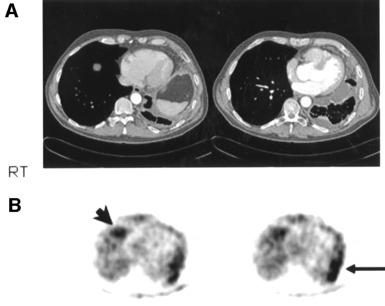
(A) Axial CT slices of thorax show circumferential pleural nodularity encasing left hemithorax with volume loss. There is medial loculation of pleural fluid, and pleural effusion is also seen in dependent location at left lung base. There is no evidence of right pleural thickening. RT = right. (B) Axial 18F-FDG-CI shows 18F-FDG hypermetabolism in left pleural cavity (arrow). In addition, there is distinct focus of increased uptake in anterior right costophrenic sulcus suggesting contralateral disease (arrowhead). Histology confirmed bilateral pleural mesothelioma of epithelial type.


