


Abstract
The purpose of this study was to assess the clinical spectrum of asymptomatic abnormal focal uptake of 99mTc-methylene diphosphonate (MDP) in the femoral neck. Methods: Fifteen consecutive patients with asymptomatic abnormal focal uptake of 99mTc-MDP in the femoral neck were evaluated. Two patients had bilateral abnormal focal uptake. The patient’s history, clinical findings, and plain hip radiograph were obtained in all cases. Scintigraphic, radiographic and clinical findings were correlated. Results: Eight of 17 (47%) femoral necks showed a definite herniation pit on radiography, 6 patients (35%) had normal radiographic findings, 1 patient had a bone island, 1 patient had a bone island and a herniation pit, and 1 patient had a subtle lesion suggestive of a herniation pit on radiography. All patients remained asymptomatic for at least a 10-mo follow-up period. Conclusion: A herniation pit is the most common finding among asymptomatic abnormal femoral neck focal uptake. This condition should be distinguished from a wide variety of disorders associated with increased focal abnormal uptake of 99mTc-MDP in the femoral neck.
Bone scintigraphy is a sensitive test that detects areas of abnormal bone turnover in various musculoskeletal disorders. Asymptomatic abnormal focal uptake of 99mTc-methylene diphosphonate (MDP) in different bones may be found in almost half of the patients who are referred for bone scintigraphy (1). Asymptomatic abnormal focal uptake in the superolateral aspect of the femoral neck is not uncommon and has been characterized as a finding of uncertain significance (2). A wide spectrum of disorders is associated with an abnormal focal uptake in the femoral neck. In this study, we assessed the clinical significance of an asymptomatic abnormal focal uptake of 99mTc-MDP in the femoral neck.
MATERIALS AND METHODS
We evaluated 15 consecutive patients (14 men, 1 woman; mean age, 23 ± 5 y; age range, 19–38 y) with an abnormal focal uptake in the femoral neck on bone scintigraphy. Eight patients were military recruits on active duty and 1 man was active in sports. They were referred for bone scintigraphy for different suspected skeletal disorders (knee injury, n = 6; lower back pain, n = 5; hand injury, n = 2; shin injury, n = 1; vasculitis or arthritis, n = 1). Whole-body radionuclide skeletal imaging was performed 2 h after intravenous injection of 925 MBq 99mTc-MDP. SPECT and pinhole images of the hips were obtained at the discretion of the interpreting physician. The patient’s history, clinical findings, and plain hip radiograph were obtained in all cases. Scintigraphic, radiographic, and clinical findings were correlated.
RESULTS
Bone scintigraphy showed a moderate focal increased uptake of 99mTc-MDP in the proximal superolateral aspect of the femoral neck in 17 femurs. All patients were asymptomatic regarding the hip region and 2 had bilateral lesions. In 8 femurs, an osteolytic lesion surrounded by a thin sclerotic border compatible with femoral neck herniation pit was found on radiography: 1 femur showed a bone island on radiography, 1 femur had 2 radiologic findings in the femoral neck (bone island and herniation pit), and 6 femurs had normal hip radiographs. In 1 case, a subtle x-ray lesion suggestive of herniation pit was found. All patients remained asymptomatic for at least a 10-mo follow-up period.
DISCUSSION
Asymptomatic abnormal focal uptake in the femoral neck may pose a clinical dilemma. In athletes or young adults who are referred for bone scintigraphy to evaluate a different symptomatic region, an abnormal focal uptake in the femoral neck may represent an asymptomatic stress fracture and should be further evaluated to prevent severe complications. The localization of the abnormal focal uptake in the femoral neck may better characterize the underlying condition causing the increased focal abnormal uptake in this region.
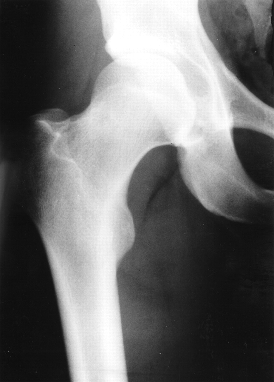
The herniation pit, first described by Pitt et al. (3) in 1982, is a subcortical bone defect located typically in the superolateral aspect of the proximal femoral neck. This benign lesion is presented histopathologically by a subcortical cavity filled with hyaline and fibrocartilaginous tissue surrounded by reactive new bone (3–5). It is seen on radiography as a relatively well-defined round or oval lucency, usually <1 cm in diameter, bordered by a thin sclerotic zone (3,4) (Fig. 1). A herniation pit has been considered a normal variant; however, the overlying cortex may fracture and cause symptoms (4).

Anteroposterior radiograph of right hip shows herniation pit of femoral neck.
A femoral neck stress fracture may progress to a complete fracture, delayed union, or nonunion and usually requires restriction of physical activity or surgical intervention (6,7). Moreover, stress fractures may not be associated with bone pain (8). Two types of femoral neck stress fractures have been described: tension and compression (9). Tension stress fractures occur typically on the distal superolateral aspect of the femoral neck, whereas compression stress fractures occur typically on the distal inferomedial aspect of the femoral neck (9,10). Tension fractures represent an increased risk for a displaced femoral neck fracture and require operative stabilization. Compression fractures are more stable and are usually treated conservatively (9). A tension fracture is more common in the elderly, and a compression fracture is more common in young adults (10).
The differential diagnosis between a herniation pit and a tension stress fracture on bone scintigraphy alone may be difficult. However, the classic scintigraphic pattern of a compression femoral neck stress fracture is a focal increased uptake in the distal inferior aspect of the femoral neck, just proximal to the lesser trochanter (11,12). According to our data and as reported in the literature (13,14), increased uptake caused by a herniation pit is located typically in the proximal superolateral aspect of the femoral neck (Figs. 2 and 3).

Schematic depiction shows focal abnormal uptake on femoral neck.

A 20-y-old soldier referred for bone scintigraphy to evaluate knee pain. Pinhole image of right hip shows focal increased uptake (arrow) corresponding to femoral neck herniation pit in same region seen on hip radiograph shown in Figure 1.
The bone island is a benign asymptomatic lesion composed of normal compact bone showing intraosseous sclerotic areas with discrete margins on radiography. Bone scintigraphy usually shows normal uptake in cases of a bone island. However, increased focal uptake can be found in cases of large bone islands or can indicate new bone formation during the beginning and growth of the lesion (15).
The usual appearance of an osteoid osteoma on bone scintigraphy is a hot spot with a focus of marked increased uptake corresponding to the site of the nidus (double-density sign), which may be located anywhere in the femoral neck (16–18). The differential diagnosis of focal abnormal uptake in the femoral neck—in addition to stress fracture, herniation pit, bone island, and osteoid osteoma—should include, in certain clinical settings, skeletal metastases and Brodie’s abscess (13,19).
We observed 5 patients with asymptomatic increased focal abnormal uptake in the femoral neck (2 with bilateral findings), who had normal radiographs. Symptoms did not develop during a follow-up period ranging from 10 to 12 mo. In these 5 patients, the scintigraphic characteristics and localization of increased radiotracer activity in the femoral neck were identical to those that showed a herniation pit on radiography. The initial phase of development or the small herniation pit possibly may be difficult to appreciate on radiography, but the bone remodeling around the subcortical cavity could be represented by an increased focal uptake of 99mTc-MDP on bone scintigraphy.
Drubach et al. (1) reported recently that asymptomatic focal abnormalities of the femur or the tibia are not commonly seen in young athletes. In this study, 5 of 8 patients, who had incidental asymptomatic focal increased uptake in the femoral neck in association with a herniation pit on radiography, were involved in intensive physical activity. These findings agree with those of previous reports, which showed that a herniation pit develops more frequently in patients with a history of intensive physical activity (4,5,20).
Among our patients with incidental increased uptake in the femoral neck, more than half showed an association between this activity of radiotracer and a herniation pit on radiography. Therefore, the combination of these 2 studies may provide the correct interpretation of this scintigraphic finding.
CONCLUSION
When incidental increased abnormal focal uptake in the femoral neck is noted on bone scintigraphy, plain radiography should be performed and a herniation pit should be included in the differential diagnosis.
Footnotes
Received Aug. 27, 2001; revision accepted Dec. 12, 2001.
For correspondence or reprints contact: David Groshar, MD, Department of Nuclear Medicine, Bnai Zion Medical Center, P.O. Box 4940, Haifa, 31048 Israel.
E-mail: d.groshar{at}b-zion.org.il
REFERENCES
In this issue


Jump to section
Related Articles
Cited By...
- No citing articles found.