


Abstract
The objective of the study was to compare relative uptake, metabolism, and β-receptor affinity of the new positron-emitting uptake-1 tracer (1R,2S)-4-18F-fluorometaraminol (4-FM) with those of the SPECT pharmaceutical meta-123I-iodobenzylguanidine (MIBG) in Wistar Kyoto (WKY) rats and spontaneously hypertensive (SHR) rats. Methods: No-carrier-added 4-18F-FM was applied to SHR and WKY rats in vivo and to retrogradely perfused hearts in vitro. Cardiac and extracardiac distribution was assessed, and metabolite formation was determined by thin-layer chromatography. The in vivo experiments were repeated with no-carrier-added 123I-MIBG. By means of autoradiography, the β-receptor affinity of 4-FM was compared with that of MIBG and propranolol (10 μmol/L) through displacement of 125I-iodocyanopindolol (1.5 pmol/L) in slices of heart and spleen. Results: Cardiomyopathic hearts showed heterogeneous 4-18F-FM uptake with gradients up to 3.6 in vivo and in vitro between different regions of the heart. Control hearts showed such gradients in 4-18F-FM uptake only in vitro. 123I-MIBG exhibited a less heterogeneous in vivo distribution in SHR hearts. Extracardiac differences between WKY and SHR were found for uptake of 4-18F-FM in the spleen (63.3% ± 4% vs. 38.8% ± 5.7% of cardiac activity) and for renal uptake of 123I-MIBG (373% ± 27% vs. 81.4% ± 17% of cardiac activity). Metabolites of 4-18F-FM were found only in the liver and those of 123I-MIBG were found in the liver and kidney with a nearly equal relative fraction in both types of animals of about 20%, 60%, and 30%, respectively. 4-FM suppressed cardiac-specific β-receptor binding of 125I-iodocyanopindolol in heart and spleen of both types of animals significantly, whereas MIBG had almost no effect. Conclusion: The more heterogeneous cardiac distribution of 4-18F-FM suggests that it reflects alterations in uptake-1 better than 123I-MIBG in addition to the possibility of quantification and higher spatial resolution by PET compared with SPECT. Altered biotransformation in cardiomyopathic diseases may also impair the evaluation of 123I-MIBG-SPECT data. The β-receptor binding of 4-18F-FM must be further elucidated.
Alterations of the adrenergic receptor population that accompany cardiomyopathic diseases are well documented experimentally and clinically (1,2). Such transformations may play a role not only in etiogenesis of hypertrophic and dilated cardiomyopathy or cardiac failure but also in patients with a predisposition to arrhythmic events (2,3).
The SPECT pharmaceutical meta-123I-iodobenzylguanidine (MIBG) has been shown to be helpful as an indicator of changes in the capacity of presynaptic norepinephrine uptake (uptake-1) in diagnosis of various tumors, such as pheochromocytoma and several types of neuroblastoma (4,5). Moreover, MIBG has been reported to identify tissue areas that are injured by metabolic diseases as well as to monitor cardiac reinnervation after cardiac transplantation surgery (6,7). However, the conclusions from the assessment of its efficacy in studies of more moderate functional impairment in cardiomyopathic or arrhythmic hearts remain contradictory (3). Nevertheless, there is no doubt that PET and SPECT radiotracers may contribute to a better understanding of the role of uptake-1 in the etiogenesis of cardiomyopathies and to an improvement of diagnostic possibilities. In addition to MIBG, several norepinephrine analogs have been labeled with short-lived nuclides with the aim of in vivo characterization of cardiac sympathetic innervation by means of PET and SPECT (8–15).
Metaraminol has been proposed as one of the first false adrenergic neurotransmitters for the measurement of presynaptic uptake-1 (16). The absence of a catechol moiety stabilizes against breakdown by catechol-O-metyltransferase. Although its affinity to vesicular storage sites has been shown to be low, the affinity to uptake-1 has been shown to exceed markedly that of norepinephrine and many other adrenergic substances (17).
11C-, 125I-, 76Br-, and 18F-labeled compounds of metaraminol (8–15,18) have been synthesized and applied in healthy rodents, monkeys, humans, and other animal experiments after chemically induced sympathetic denervation (8,10). Although radioiodinated metaraminol compounds were unsuitable for cardiac studies (15), a high biologic activity and high cardiac accumulation were observed especially for the (1R,2S)-enantiomer of 4-18F-fluorometaraminol (4-FM) and of 6-18F-fluorometaraminol (6-FM) (8,12–14). These 18F-labeled derivatives of metaraminol show rapid accumulation in the heart that remains over a period of 4 h. In comparison with 11C-metaraminol, the fluorinated derivatives of metaraminol can be assumed to have lower affinity to α-receptors in analogy to ring-fluorinated catecholamines (19). As a further alternative, the N-methyl analog of metaraminol, meta-11C-hydroxyephedrine characterized by resistance against monoamine oxidase, is often used for PET measurements (9). However, 18F tracers are not dependent on the presence of an in-house cyclotron. The specific activity achieved with no-carrier-added preparations of 4-18F-FM (14), even more than with 6-18F-FM, guarantees that PET investigations can be performed far below toxicologic doses.
This study investigates the uptake of 4-18F-FM in hearts of spontaneously hypertensive (SHR) rats and normotensive Wistar Kyoto (WKY) rats in vitro and in vivo. SHR rats develop a generally enhanced sympathetic tone and a downregulation of cardiac β1-receptors (20,21). 4-18F-FM was applied in isolated perfused hearts separated from ganglionic sympathetic regulation and in vivo with intact general sympathetic innervation to both groups of animals. A further aim of this study was to compare the regional cardiac distribution and extracardiac distribution and metabolism of 4-18F-FM and 123I-MIBG in vivo. Blocking experiments with unlabeled 4-FM and MIBG and the adrenergic radioligand 125I-iodocyanopindolol (ICYP) were performed in vitro for characterization of their respective adrenergic affinity in heart and in spleen as a further extensively sympathetic innervated organ.
MATERIALS AND METHODS
Chemicals
Most chemicals were purchased from Sigma (Deisenhofen, Germany) or Merck (Darmstadt, Germany) and were of analytic grade. 125I-(−)-ICYP (specific activity, 81.4 GBq/μmol) was supplied by NEN Life Science Products, Inc. (Zaventem, Belgium). The precursor synthesis of MIBG and its labeling were performed according to established procedures (22,23). No-carrier-added 4-18F-FM was synthesized as described (12,13). The specific activity of 4-18F-FM and 123I-MIBG applied was ≥80 GBq/μmol.
S-(−)-Propranolol, poly-(l)-lysine, and phenylmethylsufonylfluoride (PMSF) were purchased from Sigma. MIBG carbonate (22,23) and (1R,2S)-4-FM tartrate (4-FM) (12,13) were synthesized as described.
Animals
Fifty-two male SHR and WKY rats (11–13 wk old; Charles River Wiga GmbH, Sulzfeld, Germany) were used in our study. The weights of the animals were, on average, 279 ± 9 g (SHR) and 308 ± 10 g (WKY).
In Vivo Distribution Studies
Male rats (n = 11 each, WKY and SHR) were subjected to investigations in vivo. For these experiments the radiotracers were dissolved in 250–500 μL 0.9% NaCl and injected into the tail vein; 2.5 MBq/100 g body weight were used when 4-18F-FM was applied, and 3 MBq/100 g body weight were applied in 123I-MIBG experiments. The animals were anesthetized with diethyl ether and killed 30 min after injection. The hearts were excised and mounted for 2 min on a Langendorff apparatus (perfusion medium as for in vitro perfusion) for washout of tracer from vasculature and interstitium (17). Tissue samples were taken for analysis of regional distribution of the tracer from the atria, right (right ventricular basal [RVB]) and left ventricular (left ventricular basal [LVB]) regions close to the atrioventricular groove and from left (left ventricular apical [LVA]) and right ventricular (right ventricular apical [RVA]) regions close to the apex (Fig. 1). After measurement in a well-type scintillation γ-counter (Minaxi γ; Packard Instrument Co., Meriden, CT), the samples were dissolved in 0.5 mol/L NaOH containing 1% sodium deoxycholate and 0.1% sodium dodecyl sulfate to determine the protein content using a modified Lowry procedure (24).

Pattern of regions of interest to analyze cardiac distribution of 4-18F-FM and 123I-MIBG. LA = left atrium; RA = right atrium; RVB = right ventricle basal; RVA = right ventricle apical (reference region); LVB = left ventricle basal; LVA = left ventricle apical.
Any additional stress to the animals (drawing of blood samples or use of other kinds of instrumentation) that could have induced alterations in cardiac tracer uptake was avoided during the experiment. The limitations in comparing the in vivo data with the in vitro data, which were attributed to the absence of further data on hemodynamics or differences of tracer supply, were circumvented using a modification of semiquantitative methods that are common in clinical evaluation of tomographic data (6). Thus, the relative intracardiac uptake was determined using ratios of uptake in the regions of interest (ROIs) described above related to the uptake in the RVA region. The RVA region was chosen because it showed a low uptake of the tracers compared with that of other regions (average of all hearts investigated) and a small relative SD.
Ex Vivo Metabolite Studies
Ventricular heart samples were excised 30 min after injection for analysis of metabolites (frozen in liquid nitrogen until homogenization). A 30-min exposure was chosen because 4-18F-FM and 123I-MIBG show an accumulation in the heart that is well detectable at this time after injection and MIBG is known to have a high metabolic activity (25). Besides the cardiac samples, tissue samples of kidney, liver, spleen, and skeletal muscle were subjected to metabolite analysis. The samples were homogenized with a microhomogenizer in 3 volumes of 0.5 mol/L ice-cold perchloric acid and centrifuged at 12,000g for 10 min at 0°C. The supernatant was neutralized with 5 mol/L K2CO3 and used for thin-layer chromatographic (TLC) analysis of metabolites after a second centrifugation for removal of potassium perchlorate. TLC was performed with 5–10 μL of the neutralized supernatant using Si-60-F254 foils (Merck, Darmstadt, Germany) and 1,4-dioxane/isobutanol/triethylamine/water (1:1:1:1, vol/vol) as eluent. Separation of metabolites of 123I-MIBG was performed with RP-18 F254S foils (Merck) and acetonitrile/0.01 mol/L NaH2PO3 (4:96, vol/vol) as eluent. Measurement of the radioactivity of the TLC plates was performed using an InstantImager (Packard Instrument).
In Vitro Perfusion Studies
From SHR and WKY male rats (n = 5 each), hearts were excised after anesthesia with diethyl ether, cervical dislocation, and midline thoracotomy. The hearts were rinsed briefly with ice-cold saline and mounted on a Langendorff apparatus (homemade) within 30–60 s, starting perfusion immediately through an aortic cannula. The perfusion medium contained 118 mmol/L NaCl, 25 mmol/L NaHCO3, 2.9 mmol/L KCl, 1.18 mmol/L MgSO4, 1.28 mmol/L KH2PO4, 1.6 mmol/L CaCl2, and 11 mmol/L glucose supplied with a mixture of 95% O2 and 5% CO2 (pH 7.4, 37°C). Preparation of pairs of WKY and SHR hearts was done in the same way in parallel. 4-18F-FM supplied from the same batch was used for simultaneous perfusion in each experiment.
After an equilibration period of 10–15 min, 3.7 MBq 4-18F-FM were added to the perfusion medium within 10 min (0.5 mL/min) using a slow-velocity infusion pump (Braun, Secura Melsungen, Germany) at a total coronary flow of 9 ± 0.8 mL/min and an aortic pressure of 60 mm Hg. The infusion of the tracer was followed by a 2-min washout with perfusion medium as described (17). The hearts then were removed immediately from the perfusion apparatus and dried gently on filter paper. Samples were excised from different heart regions as described above for measurement of distribution. The tissue samples were placed in scintillation cuvettes, and radioactivity was determined in a Minaxi γ-counter.
In Vitro Receptor Studies
Blocking of 125I-ICYP binding was performed in vitro with tissue of further groups of animals (male WKY and SHR; n = 5 each) to compare the β-adrenergic receptor affinity of 4-FM, MIBG, and S-(−)-propranolol. For this purpose, the hearts and spleens of WKY and SHR rats were frozen in isopentane at −50°C and kept at −70°C.
The excised organs were cut 1 d later in a CM3050 cryostat microtome (Leica, Nussloch, Germany). Consecutive sections were immediately mounted on microscopic slides coated with poly-(l)-lysine. The incubation procedure was performed with a slight modification of the procedure in the literature (26). The slices were preincubated for 15 min at room temperature in a buffer (pH 7.6) containing 170 mmol/L Trizma, 5 mmol/L MgCl2, 0.01% ascorbic acid, and 0.25 mmol/L PMFS (end concentration after dilution of a 165 mmol/L stock solution in absolute ethanol in the buffer). The incubation was performed for 120 min at room temperature with 1.5 pmol/L 125I-ICYP. Control samples and samples blocked with S-(−)-propranolol, 4-FM, or MIBG (10 μmol/L each) were coincubated in the same experiment.
The slices were incubated subsequently for 2 × 5 min in ice-cold Tris-buffered medium described above for removal of free 125I tracer. They then were rinsed briefly in ice-cold distilled water and dried in a stream of air. An imaging plate (BAS-SR; Fuji Film, Straubenhardt, Germany) was exposed to the samples for 24 h. Analysis of photostimulated luminescence was performed with a BAS 5000 bioimage analyzer (Fuji Film).
Statistics
Statistical analysis of the data from the in vitro experiments was done by 1-way ANOVA and the Bonferroni parametric t test for multiple comparisons. Statistical analysis of the data obtained in the in vivo experiments was performed using the nonparametric Kruskal–Wallis test and paired t test. P < 0.05 was taken as limit of statistical significance.
RESULTS
Cardiac Uptake of 4-18F-FM and 123I-MIBG
Evaluation of the cardiac distribution was based on the uptake of 4-18F-FM and 123I-MIBG measured directly in samples of different ROIs (Fig. 1) and related to the protein content of the respective tissue. The in vivo uptake of the 2 compounds measured directly amounted to 5–50 cpm/μg protein in different ROIs, whereas the uptake in the in vitro perfusion experiments was 5–10 times higher than that in the respective regions in vivo. In the reference region (RVA), after injection of 4-18F-FM in vivo, 10.0 ± 2.26 and 5.7 ± 1.5 cpm/μg protein were found in WKY and SHR hearts, respectively. The respective values of the in vitro experiments were 75.46 ± 11.1 cpm/μg protein for WKY hearts and 59.47 ± 8.3 cpm/μg protein for SHR hearts. After application of 123I-MIBG in vivo, the uptake in the RVA of WKY hearts was 11.5 ± 1.76 cpm/μg protein and that of SHR hearts was 12.5 ± 1.74 cpm/μg protein. To allow a direct comparison between cardiac distributions of 123I-MIBG- and 4-18F-FM, data of the in vivo experiments were corrected for differences in the applied dose and physical nuclide decay during the 30-min exposure.
The relative uptake values given in Tables 1 and 2 were obtained by relating the uptake in the respective ROIs with the uptake of the RVA region as a common reference region. Evaluation of the in vivo ratios (ROI/RVA) after injection of 4-18F-FM and, to a lesser extent, of 123I-MIBG reveals differences between healthy WKY and diseased SHR hearts (Table 1), with pronounced intracardiac alterations in comparison with control hearts after application of 4-18F-FM. Whereas ratios of 4-18F-FM uptake were found in the range of 1.1–1.5 for WKY hearts, hearts of SHR animals showed ROI/RVA of 1.5–3.6. In contrast, maximum gradients of uptake of 123I-MIBG in diseased animals in vivo were <2. Differences of uptake ratios of the tracers between WKY and SHR hearts were most significant in the right ventricle for both: 4-18F-FM (WKY, 1.29 ± 0.26; SHR, 2.98 ± 0.6; P < 0.05) and 123I-MIBG (WKY, 0.97 ± 0.04; SHR, 1.27 ± 0.09; P < 0.05). In contrast, the perfusion with 4-18F-FM in vitro resulted in a heterogeneous distribution in WKY and SHR hearts. The highest ROI/RVA in WKY hearts was 2.5 in the LVB region and the lowest was 1.03 in the atrium. The respective values for SHR were 3.6 and 0.99 (Table 2).
Relative Uptake of 4-18F-FM and 123I-MIBG in SHR and WKY Rats In Vivo
Relative Uptake of 4-18F-FM in Retrogradely Perfused Hearts of SHR and WKY Rats In Vitro
Relative Uptake and Ex Vivo Metabolite Studies
After application of 4-18F-FM, metabolites were found only in the liver of WKY and SHR animals (Fig. 2). No metabolites were detected in the heart or any other organ examined. The relative metabolite fraction was similar in the liver of WKY and SHR animals (20.6% ± 1.8% vs. 22.6% ± 7% of total liver activity).

Relative uptake and metabolites of 4-18F-FM in SHR and WKY tissue samples. Total uptake in tissue of organs and in blood is given as percentage of cardiac activity (mean ± SEM). Black columns, SHR (n = 3); white columns, WKY (n = 3). Hatched areas (mean ± SEM) show contribution of metabolites to total uptake. Asterisk indicates significant difference between SHR and WKY animals.
The uptake of 4-18F-FM in spleens of WKY rats was higher than that in spleens of SHR rats (63.36% ± 4.1% vs. 38.8% ± 5.7% of cardiac activity; P < 0.05) but, in turn, was slightly lower than that in skeletal muscle of SHR (7.16% ± 0.68% vs. 10.9% ± 0.35% of cardiac activity). No significant differences could be seen in liver (WKY, 109.5% ± 32% of cardiac activity; SHR, 116% ± 8.5%), kidney (WKY, 38.2% ± 6.3%; SHR, 51.4% ± 17.1% of cardiac activity), or blood (7.56% ± 1% vs. 7.4% ± 0.9% of cardiac activity).
TLC separation of tissue extracts after injection of 123I-MIBG revealed significant differences in general uptake of the tracer and of the activity fraction caused by metabolites between WKY and SHR only in the kidney (Fig. 3 [also shown as percentage of cardiac activity]). Thus, the total activity in kidney samples of WKY was nearly 5-fold that of SHR (373% ± 27% vs. 81.4% ± 17% of cardiac activity; P < 0.05; n = 6 each). In contrast, differences in uptake were negligible in spleen and liver. For WKY the renal metabolite fraction corresponds to 123.2% ± 43.5% and for SHR this fraction corresponds to 24% ± 6% of cardiac activity (P < 0.05). Thus, metabolites reached values of 30% ± 9.7% and 29% ± 3.6% of total renal activity, respectively. The percentage of 123I-MIBG breakdown products of total activity in the liver was approximately 3-fold of that of 4-18F-FM in both WKY and SHR (58.7% ± 5.8% and 64.5% ± 6.8% of total liver activity). However, in spite of the differences between the radiotracers, the relative fraction of metabolites in the respective tissue was always nearly equal for both types of animals.

Relative uptake and metabolites of 123I-MIBG in SHR and WKY tissue samples. Total uptake in tissue of organs and in blood is given as percentage of cardiac activity (mean ± SEM). Black columns, SHR (n = 6); white columns, WKY (n = 6). Hatched areas (mean ± SEM) show contribution of metabolites to total uptake. Asterisks indicate significant difference between SHR and WKY animals.
In Vitro Receptor Studies
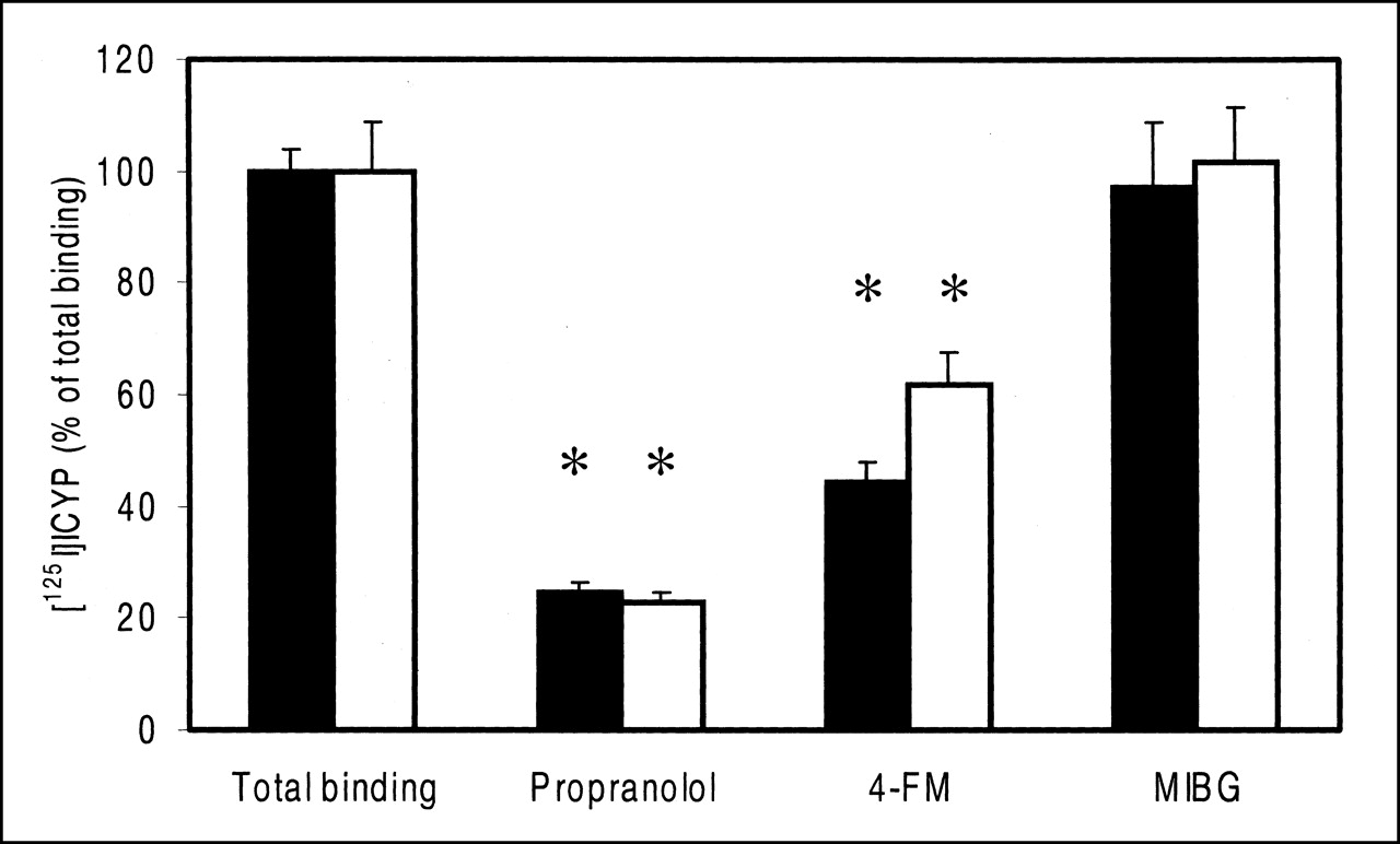
The nonspecific binding of 125I-ICYP, determined by displacement with the β-receptor antagonist S-(−)-propranolol, was markedly higher in cardiac sections (Fig. 4) than in spleen slices (Fig. 5); 57.7% ± 4.3% of the total binding remained after exposure to S-(−)-propranolol in WKY cardiac samples and 66.7% ± 5% in SHR cardiac samples. 4-FM decreased 125I-ICYP binding in the hearts of healthy animals to 65.82% ± 7.6% of total binding (P < 0.05), whereas MIBG was without any significant effect. Neither 4-FM (86.9% ± 8.9% of total binding) nor MIBG altered the binding of 125I-ICYP in cardiac sections of SHR significantly.

Effect of S-(−)-propranolol, MIBG, and 4-FM on binding of 123I-ICYP in cardiac tissue of SHR and WKY animals. Inhibitory efficiency of β-receptor binding of 4-FM is compared with that of MIBG and propranolol (125I-ICYP, 1.5 pmol/L; inhibitors, 10 μmol/L each). Black columns, SHR (n = 6–8); white columns, WKY (n = 6–8). Data are expressed as mean ± SEM. Asterisks indicate significant differences to total binding.
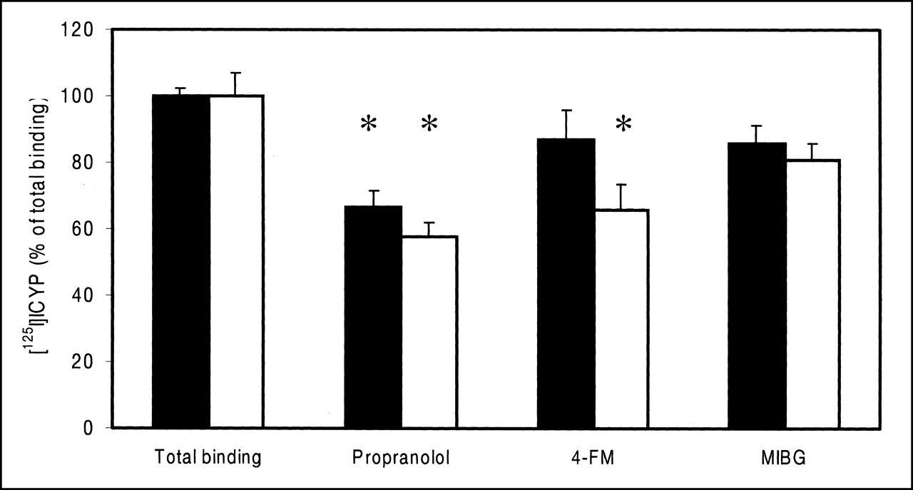
Effect of S-(−)-propranolol, MIBG, and 4-FM on binding of 125I-ICYP in spleen of SHR and WKY animals. Inhibitory efficiency of β-receptor binding of 4-FM is compared with that of MIBG and propranolol (125I-ICYP, 1.5 pmol/L; inhibitors, 10 μmol/L each). Black columns, SHR (n = 11); white columns, WKY (n = 11). Data are expressed as mean ± SEM. Asterisks indicate significant differences to total binding.
In general, spleen slices showed twice the total radioligand binding observed in the hearts. Nonspecific binding after exposure to S-(−)-propranolol in WKY spleen sections was 22.9% ± 1.77% of total binding and 24.8% ± 1.67% in SHR spleen sections (Fig. 5). Application of 4-FM to WKY spleen sections decreased the percentage of binding of 125I-ICYP to 61.76% ± 6% and had an even larger effect on radioligand binding in SHR slices (44.2% ± 3.6% of total binding; P < 0.05), whereas MIBG had no influence on the binding of the radioligand.
DISCUSSION
Previous investigations on the cardiac uptake of the SPECT pharmaceutical 123I-MIBG and the potential PET tracer 4-18F-FM revealed a somewhat different distribution of these tracers in the presynaptic compartment and also differences in pre- and postsynaptic monoamine transport (14,27). To our knowledge, the consequences of these differences on cardiac uptake have not been investigated in a pathophysiologic model. This analysis of the cardiac uptake of 4-18F-FM in vitro and in vivo revealed 2 noteworthy observations: low uptake in the atria compared with that in the ventricles and a heterogeneous uptake in SHR rats in vitro and in vivo but absence of intracardiac gradients of tracer accumulation in normotensive WKY animals in vivo.
The relatively low atrial 4-18F-FM content in SHR and WKY hearts in vitro as well as in WKY hearts in vivo and also with 123I-MIBG in vivo is, on a first glance, in contradiction to former histofluorescence (28) and biochemical studies (29). Atrial sympathetic innervation and endogenous norepinephrine content in atria are twice the values of the ventricles (29). However, recently there is increasing evidence for an essential role of β2-receptors in the heart, including presynaptic activation of norepinephrine release (30). The heterogeneity of tracer uptake in SHR animals compared with WKY animals, which became evident during analysis of relative 4-18F-FM uptake, can be discussed with respect to coronary local hemodynamics and to differences in regulation of cardiac sympathicus as well. SHR animals are well known to develop alterations in systolic and diastolic pressure as well as mean arterial pressure already at the age of the rats used (11–13 wk) (31). The total coronary flow, capillary density of the heart, and coronary function of SHR animals do not differ from those of control animals during the first 22 wk without additional physical exercise (21,31). Accordingly, in our experiments no differences were found in total coronary flow between SHR and WKY animals. On the other hand, it is well known that differences of the norepinephrine content of extracardiac organs, such as kidney, occur in SHR rats early in life (32). The role of alterations of the supracardiac receptor population for cardiac norepinephrine turnover, the density or activity of uptake-1 transporters, and the perfusion of cardiac microcompartments have not been clarified; thus, their influence remains open.
Results after application of MIBG are in accordance with the report of Matsunari et al. (25), which described only transient alterations between uptake in the endocardium and epicardium during the first 3 h after tracer application but marked permanent differences 6 h after injection of MIBG during the plateau phase of the time–activity curve (25). Differences in the mechanisms of uptake of the 2 tracers may play an essential role for the divergent distributions we found with 4-18F-FM and 123I-MIBG in SHR and WKY rats in vivo. Two mechanisms are known for the presynaptic uptake of MIBG. One, a neuronal mechanism, is a specific, saturable, energy-dependent mechanism (27,33–35). Na+/Cl− gradients have been postulated to drive this uptake mediated by the Na+,K+-adenosinetriphosphatase. The second, an extraneuronal uptake mechanism, is energy independent, unsaturable, and nonspecific (27).
Reports of 0%–94% inhibition of uptake-1 of MIBG vary depending on the model used (27,35) and show an extravesicular portion of 50% of presynaptic tracer accumulation after application of reserpine. In isolated working rat hearts, desipramine (50 nmol/L) suppressed only 27% of the neuronal MIBG uptake, and a high contribution of uptake-2 to the extraneuronal uptake was shown after additional application of the uptake-2 inhibitor SKF 550 (27). In contrast, application of reserpine resulted in a 76% inhibition of 4-18F-FM uptake in the left ventricle of rats (14), suggesting that the fraction of MIBG in the extravesicular compartment is larger than that of 4-18F-FM.
In addition to the cardiac response, the spleens of SHR rats showed a lower 4-18F-FM uptake than that of WKY animals. For 123I-MIBG, a decreased accumulation has been described in clinical studies on spleens of patients with congestive cardiomyopathy (36) but not in experiments using rats (32). The heart and also the spleen are known to have a high density of β1-receptors as well as a presynaptic population of β2-receptors (30,37) mediating norepinephrine release. Whether such mechanisms are causal for the differences in uptake of 4-18F-FM in spleens of rats or of 123I-MIBG in spleens of humans remains to be elucidated.
Despite their differences in cardiac distribution, 4-FM and MIBG are similar in their stability against the neurojunctionally localized enzymes monoamine oxidase and catechol-O-methyltransferase, key enzymes in the catabolism of catecholamines. This was an essential reason for the choice of these radiotracers as potential diagnostic tools. The absence of the hydroxyl group in the 4-position and the presence of the methyl group in the α-position in metaraminol ensure resistance against the 2 enzymes. Possibly, as a consequence of these properties, metabolites were not found in the heart with either 4-18F-FM or with 123I-MIBG. Nevertheless, breakdown products of metaraminol and 6-18F-FM have been described in biodistribution studies (9,10,38). It was assumed that metaraminol could be hydroxylated in the 4-position, resulting in α-methylnorepinephrine, and subsequently 3-O-methylated to α-methyl-normetanephrine (11,38). In these studies, 4-18F-FM has been assumed to show further restriction against formation of metabolites because metabolism occurs predominantly in the 3- and 4-positions (11). Thus, our data with 4-18F-FM (aside from a minor fraction of 20% of activity in the liver) seem to confirm these previous suggestions. Most data on the metabolism of the parent compound metaraminol indicate a very slow breakdown of metaraminol in rats and a rapid metabolism in guinea pigs (31,38). However, these data were obtained after intraperitoneal injection (38), which is not comparable with the model used in this study. A single study describes a rapid decrease of 11C-metaraminol and the occurrence of labeled metabolites in the plasma of a monkey. However, the study did not provide further identification of the metabolites (12).
For 123I-MIBG, we observed a larger breakdown in the liver and kidney 30 min after injection, which is in accordance with previous reports (25,39). In this study, approximately 60%–65% of liver activity and nearly 30% of kidney activity were metabolites. Kidneys of WKY animals accumulated 3.7-fold higher levels of metabolites than the levels in the heart, but kidneys of SHR animals accumulated only 0.81-fold. Our data, with only 1 time point of biotransformation, permit no final conclusion on whether differences in the time course or in the enzymatic pattern between SHR and WKY rats are responsible for these alterations. Neither 4-18F-FM nor 123I-MIBG showed metabolites in the heart at the time of investigation. For 4-18F-FM, this is favorable compared with 6-18F-FM, for which Rosenspire et al. (10) described metabolic products in the heart.
The investigation of the β-receptor affinity of MIBG and 4-FM in the heart and spleen revealed different patterns for SHR and WKY tissue and for the 2 agonists as well. The β-adrenergic radioligand 125I-ICYP showed a 3-fold higher nonspecific binding in heart slices than that in the spleen in both SHR and WKY. 4-FM suppressed 76.1% ± 15% of the β-receptor–specific binding in the hearts of healthy animals. This extent is typical for the proportion of β1-receptors in normal hearts. Comparison with the weak effect of 4-FM in SHR hearts, where the number of β1-receptors is small (1), may indicate a relatively specific affinity to β1-receptors. β1-Adrenergic affinity has been described for ring-fluorinated epinephrines (19). Thus, a β1-adrenergic agonistic activity similar to that of epinephrine was described for 2-fluoronorepinephrine (19), whereas 6-fluoroepinephrine was an agonist for α-adrenoreceptors. In contrast to cardiac slices, the reduction of 125I-ICYP binding in spleen slices by 4-FM was higher in SHR than in WKY animals. 4-FM displaced 74.1% ± 5% of specific binding in SHR and 50% ± 7.9% in WKY slices. In the case of a subtype specificity of 4-FM, this would mean that cardiomyopathic disease is accompanied by an increase of β1-receptors in the spleen but not in the heart.
In contrast to the derivative of metaraminol, application of MIBG to heart slices reduced the specific binding of 125I-ICYP by only approximately 40% (no statistical significance). Reduction of MIBG uptake in the heart by the β-blocking drugs metoprolol and labetolol was observed in application of extremely high doses (>1 mmol/L) (40). Contrary to these experiments, our data do not reflect the interaction between adrenergic receptors and MIBG uptake but only differences in receptor affinity between the drugs. In contrast to 4-FM, there was no significant effect of MIBG on 125I-ICYP binding. Thus, the absence of adrenergic receptor affinity and relatively low affinity to uptake-1 of MIBG must be balanced against some adrenergic affinity of 4-FM and high affinity to uptake-1.
CONCLUSION
4-18F-FM is a potential PET tracer that is able to reflect cardiac and supracardiac disturbances of sympathetic regulatory mechanisms, and also of other intensively sympathetic innervated organs such as the spleen, more markedly than the SPECT tracer 123I-MIBG. 4-18F-FM may be considered as an alternative to 6-18F-FM and 123I-MIBG in visualization of cardiac alterations in uptake-1 because of the availability of the tracer with a high specific activity, a much lower rate of metabolization with less response to disease-related alterations in biotransformation, and a higher specificity for alterations in uptake-1. It remains to be elucidated whether the relatively high affinity to β-receptors, which is different from that of MIBG, attenuates these advantages of the PET tracer.
Acknowledgments
The authors thank Dr. Kurt Hamacher for expert advice and Erika Wabbals and Bettina Palm for the preparation of no-carrier-added 4-18F-FM.
Footnotes
Received Apr. 23, 2001; revision accepted Nov. 12, 2001.
For correspondence or reprints contact: Margit Pissarek, MD, Institut für Nuklearchemie, Forschungszentrum Jülich GmbH, Leo-Brandt-Strasse, Jülich, D-52425, Germany.
E-mail: m.pissarek{at}fz-juelich.de
REFERENCES
In this issue


Jump to section
Related Articles
Cited By...
- No citing articles found.