


Abstract
The effectiveness of 11C-choline PET in detecting various cancers, including prostate cancer, is well established. This study was aimed at developing an 18F-substituted choline analog, 18F-fluoroethylcholine (FECh), as a tracer of cancer detection. Methods: No-carrier-added 18F-FECh was synthesized by 2-step reactions: First, tetrabutylammonium (TBA) 18F-fluoride was reacted with 1,2-bis(tosyloxy)ethane to yield 2-18F-fluoroethyl tosylate; and second, 2-18F-fluoroethyl tosylate was reacted with N,N-dimethylethanolamine to yield 18F-FECh, which was then purified by chromatography. An automated apparatus was constructed for preparation of the 18F-FECh injection solution. In vitro experiments were performed to examine the uptake of 18F-FECh in Ehrlich ascites tumor cells, and the metabolites were analyzed by solvent extraction followed by various kinds of chromatography. Clinical studies of 18F-FECh PET were performed on patients with untreated primary prostate cancer as follows: A dynamic 18F-FECh PET study was performed on 1 patient and static PET studies were performed on 16 patients, and the data were compared with those of 11C-choline PET on the same patients. Results: 18F-FECh was prepared in high yield and purity. The performance of the automated apparatus was excellent. The in vitro experiment revealed that 18F-FECh was incorporated into tumor cells by active transport, then phosphorylated (yielding phosphoryl-18F-FECh) in the cells, and finally integrated into phospholipids. The clinical PET studies showed marked uptake of 18F-FECh in prostate cancer. A dynamic PET study on 1 patient revealed that the blood level of 18F-FECh decreased rapidly (in 1 min), the prostate cancer level became almost maximal in a short period (1.5 min) and it remained constant for a long time (60 min), and the urinary radioactivity became prominent after a short time lag (5 min). Static PET studies conducted under bladder irrigation showed no difference between 18F-FECh uptake and 11C-choline uptake in prostate cancer. However, 18F-FECh gave a slightly higher spatial resolution of the image, which was attributed to the shorter positron range of 18F. Conclusion: The synthesis of 18F-FECh was easy and reliable. 18F-FECh PET was very effective in detecting prostate cancer in patients. The chemical trap, consisting of active transport of 18F-FECh and formation of phosphoryl-18F-FECh, seemed to be involved in the uptake mechanism of 18F-FECh in tumors.
In most cancers a high content of phosphorylcholine has been revealed by 31P nuclear magnetic resonance (NMR) studies, whereas in the corresponding normal tissues phosphorylcholine is present at low levels, occasionally below detection (1,2). Phosphorylcholine, a product of the choline kinase reaction, is the first intermediate in the stepwise incorporation of choline, (CH3)3N+CH2CH2OH, into phospholipids by the Kennedy pathway. Katz-Brull and Degani (3) investigated choline transport in human breast cancer cells in vitro by 31P, 13C, and 2H NMR and found that choline was incorporated into the tumor cells by a carrier-mediated mechanism and then it was converted into phosphorylcholine within 1 h. Haeffner (4) investigated choline transport in Ehrlich ascites tumor cells using 3H-choline and 14C-choline. When choline was incubated with tumor cells at a low concentration, it was incorporated into the cells by an active-transport mechanism, then it was converted into phosphorylcholine also within 1 h, and finally it was integrated into phosphatidylcholine.
We previously developed 11C-choline as a PET tracer for cancer detection and have succeeded in visualizing brain tumor (5), lung cancer (6), esophageal cancer (7), colon cancer (8), bladder cancer (8), prostate cancer (9), and many other cancers (8). Motivated by this success, we attempted to develop an 18F-labeled choline analog as a PET tracer, with an idea that 18F labeling would be superior to 11C labeling because of the longer half-life and the shorter positron range of 18F. We thought that 18F-fluoroethylcholine (FECh) would be appropriate for this purpose. The following evidence supports our idea: Deves and Krupka (10) studied the binding affinity of the choline transport system for synthetic choline analogs, using red blood cells, and found that 2 methyl groups were essential, but the third methyl group was replaceable with a longer alkyl group. Clary et al. (11) studied the substrate specificity of choline kinase for synthetic choline analogs, using yeast choline kinase, and found that the 2 methyl groups and the hydroxylethyl side chain were essential, but the third methyl group was replaceable with a longer alkyl group. We had already synthesized 18F-FECh and studied its biodistribution in normal and tumor-bearing rabbits; our results are reported in a preliminary form (12). In this article, we report the details of the synthesis, biochemistry, and clinical application of this compound.
MATERIALS AND METHODS
Synthesis of 18F-FECh
We first synthesized nonradioactive FECh (fluoroethanol method) and then used it as standard for the synthesis of radioactive (no-carrier-added) 18F-FECh (tetrabutylammonium [TBA] method) (Fig. 1).

Scheme of synthesis of FECh and 18F-FECh. Fluoroethanol method produces nonradioactive FECh tosylate as final product. TBA method produces radioactive 18F-FECh tosylate as final product.
Reagents were purchased from Sigma-Aldrich Japan (Tokyo, Japan), Merck Japan (Tokyo, Japan), Wako (Osaka, Japan), Tokyo Kasei (Tokyo, Japan), YMC Co. (Kyoto, Japan), Nihon Waters (Tokyo, Japan), or Dionex Japan (Tokyo, Japan). 1H NMR (300 MHz) was measured with an NMR spectrometer (JNM-AL300 FT; JEOL, Tokyo, Japan). The mass spectrum (MS) was measured with a mass spectrometer (GCMS-QP5050; Shimadzu, Kyoto, Japan). High-resolution mass spectrometry was performed by Daikin Industries (Tsukuba, Japan). The infrared (IR) spectrum was measured with an IR spectrometer (FTIR-8300; Shimadzu). The quantitative analysis of FECh was performed by ion chromatography (Dionex DX-120; Dionex Japan). 18F-Fluoride anion was produced by proton irradiation of 18O-water using a cyclotron (Baby Cyclotron 2010N; Japan Steel Works, Tokyo, Japan).
2-Fluoroethyl Tosylate (Fluoroethanol Method).
At 0°C while stirring in an argon atmosphere, 2-fluoroethanol (1.92 g, 30 mmol) and tosyl chloride (6.87 g, 36 mmol) were dissolved in dichloromethane (30 mL), and pyridine (10 mL) and 4-dimethylaminopyridine (100 mg) were added. After remaining at room temperature for 3 d, the mixture was diluted with dichloromethane (100 mL) and then washed with 5% HCl (3 times) and brine. It was dried over MgSO4 and evaporated in vacuo to give an oil. Thin-layer chromatography (TLC) (silica gel; n-hexane:ethyl acetate, 3:1) of the oil showed 3 reaction products: vinyl tosylate (Rf, 0.57), 2-fluoroethyl tosylate (Rf, 0.36), and 1,2-bis(tosyloxy)ethane (Rf, 0.21). The product was purified by column chromatography (silica gel; n-hexane:ethyl acetate, 5:1) to give 2-fluoroethyl tosylate as a colorless oil (4.4 g, 67%). 1H NMR (CDCl3): δ 2.46 (s, 3H), 4.28 (dt, J = 27.3, 4.0 Hz, 2H), 4.56 (dt, J = 47.0, 4.0 Hz, 2H), 7.37 (d, J = 7.9 Hz, 2H), 7.82 (d, J = 7.9 Hz, 2H). MS: m/z 218, 172, 155, and 91. High-resolution MS: calculated for C9H11FSO3, 218.041; observed, 218.042. The product was analyzed by high-performance liquid chromatography (HPLC): Column, ODS-silica gel (ODS-A; YMC), 250 × 10 mm; solvent, 50 mmol/L phosphoric acid + 1 mmol/L 2-naphthalenesulfonic acid; flow rate, 5 mL/min; detector, refractometer. The retention time of the product was 3.9 min.
FECh Tosylate (Fluoroethanol Method).
2-Fluoroethyl tosylate (996 mg, 4.56 mmol) was dissolved in N,N-dimethylethanolamine (407 mg, 4.56 mmol) and, under argon, heated at 100°C for 10 min. The resultant syrup was dissolved in methyl acetate:methanol (10:1), and the solvent was removed by evaporation to give the product as colorless rhomboid crystals. After recrystallization from the same solvent, mp 63.9°C–64.5°C (1.43 g, 100%). 1H NMR (CD3OD): δ 2.37 (s, 3H), 3.23 (s, 6H), 3.54–3.61 (m, 2H), 3.76–4.04 (m, 4H), 4.90 (bd, J = 48.0 Hz, 2H), 7.23 (d, J = 8.4 Hz, 2H), 7.71 (d, J = 8.4 Hz, 2H). IR (KBr): 3,375, 2,976, 1,478, 1,213, 1,195, 1,124, 1,035, 1,012, and 684 cm−1.
FECh Chloride (Fluoroethanol Method).
FECh tosylate (50 mg, 0.16 mmol) was dissolved in methanol (20 mL) and passed through an anion-exchange resin, Amberlite IRA-900 (Cl−1) (Sigma, St. Louis, MO) (3 g). Removal of methanol gave FECh chloride as colorless needle crystals (28 mg, 100%). 1H NMR (CD3OD): δ 3.27 (s, 6H), 3.58–3.65 (m, 2H), 3.82–4.07 (m, 4H), 4.95 (bd, J = 50.0 Hz, 2H). IR (KBr): 3,383, 3,020, 1,475, 1,082, 957, 931, and 689 cm−1.
FECh Hydroxide (Fluoroethanol Method).
FECh tosylate (40 mg, 0.13 mmol) was dissolved in water (30 mL) and passed through an anion-exchange resin, Amberlite IRA-900 (OH−1) (2 g). Removal of water gave FECh hydroxide as a colorless oil (18 mg, 92%). 1H NMR (CD3OD): δ 3.26 (s, 6H), 3.55–3.69 (m, 2H), 3.78–4.07 (m, 4H), 4.98 (bd, J = 50.0 Hz, 2H). IR (KBr): 3,444, 1,474, 1,387, 1,350, 1,084, 1,052, and 957 cm−1. On the ODS-silica gel HPLC performed under the same condition as above, the retention time of FECh hydroxide was 4.6 min.
2-18F-Fluoroethyl Tosylate (TBA Method).
No-carrier-added 18F-fluoride (approximately 370 MBq), collected from an anion-exchange cartridge by elution with 2 mL 40 mmol/L TBA bicarbonate in acetonitrile:water (4:1), was dried by evaporation at 100°C and dried again with 2 mL dry acetonitrile. After addition of 1,2-bis(tosyloxy)ethane (20 mg) in dry acetonitrile (1 mL), the mixture was heated at 80°C for 20 min. After the solvent was evaporated at 80°C under reduced pressure, the dry residue was analyzed on TLC and HPLC. The Rf on TLC was identical with that of 2-fluoroethyl tosylate from the fluoroethanol method. On HPLC, the retention time of the product was identical with that of 2-fluoroethyl tosylate. A small amount of radioactivity remained in the column head.
18F-FECh Hydroxide (TBA Method).
No-carrier-added 2-18F-fluoroethyl tosylate prepared as above was dried and then dissolved in N,N-dimethylethanolamine (0.3 mL). The mixture was heated at 100°C for 5 min. After evaporation of N,N-dimethylethanolamine at 100°C under high vacuum, the dry residue was analyzed by HPLC. On the ODS-silica gel HPLC, a single radioactive peak, corresponding to 18F-FECh hydroxide, was found at 4.6 min. There was no radioactive peak of 2-18F-fluoroethyl tosylate. The radiochemical yield of 18F-FECh hydroxide compared with 18F-fluoride was 46.3% (decay corrected). The rest of the radioactivity was found in the reaction vessel and the column head. On this HPLC, a sharp mass peak of TBA (detected by a refractometer) appeared far behind the radioactive peak of 18F-FECh hydroxide. There was no other mass peak that eluted closely to 18F-FECh hydroxide.
Automated Synthesis of No-Carrier-Added 18F-FECh Chloride
Design of Automated Apparatus.
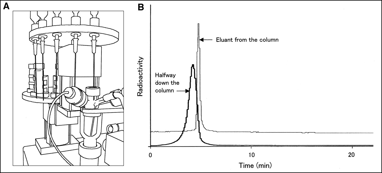
We constructed an automated apparatus for the synthesis of 18F-FECh. The function of this apparatus was as follows: (a) transfer of proton-irradiated 18O-water into a transit vessel and separation of 18F-fluoride ions using an ion-exchange cartridge, (b) transfer of 18F-fluoride ions into a reaction vessel, (c) 2-step chemical reactions (2-18F-fluoroethyl tosylate synthesis followed by 18F-FECh synthesis) in the reaction vessel, (d) passage of the product through anion-exchange cartridges to remove anionic by-products, (e) transfer of the effluent from cartridges to an HPLC apparatus, (f) HPLC, (g) passage of the effluent through anion-exchange cartridges to remove anionic molecular species in the HPLC solvent (phosphoric acid and 2-naphthalenesulfonic acid), (h) passage of the effluent from the cartridges through a cation-exchange cartridge to trap 18F-FECh, (i) washing of the cartridge with water, and (j) elution of 18F-FECh from the cartridge with saline. A close-up illustration of the reaction vessel is shown in Figure 2A, where the top of the vessel is sealed by a rubber septum, and 3 side arms are fixed on the side of the vessel. All chemical reactions were performed in this reaction vessel. Delivery of reagents to the reaction vessel was achieved by moving the upper and lower turntables; the upper table was equipped with needle-and-syringe units, and the lower table was equipped with reagent reservoirs. After one of the reagents was sucked from a reservoir into the corresponding needle-and-syringe unit, the upper and lower turntables moved vertically and rotationally, and the reagent was injected then into the reaction vessel from the needle pierced through the rubber septum. The purpose of the side arms was (a) to receive 18F-fluoride ions into the vessel; (b) to create negative, neutral, and positive pressures within the vessel; and (c) to send helium gas to the bottom of the vessel or transport the product of synthesis to the next HPLC unit. The reaction vessel could be heated by blowing hot air toward the bottom or toward the top and bottom simultaneously. The reaction vessel could be cooled by blowing chilled air. The temperature, pressure, and radioactivity of the reaction vessel were displayed continuously on a computer screen. A charged-coupled device camera and a monitor screen were used for the display of an actual view of the reaction vessel.

(A) Automated apparatus for 18F-FECh synthesis. Reaction vessel is shown close up. Purification module is out of view. (B) Preparative HPLC of 18F-FECh in automated apparatus for 18F-FECh synthesis. Column used was ODS-silica gel column, 250 × 10 mm; solvent, 50 mmol/L phosphoric acid + 1 mmol/L 2-naphthalenesulfonic acid; flow rate, 5 mL/min. Radioactivity “halfway down the column” is reading of detector (detector 1) placed on side of column to watch approach of 18F-FECh. Radioactivity of effluent from column was monitored by another detector (detector 2).
Operation of Automated Apparatus.
The routine production of 18F-FECh injection solution was performed as follows. 18F-HF was produced by the irradiation of 60% enriched 18O-water with the 20-MeV proton beam at 30 μA for 30 min. After the end of bombardment, the 18F-HF solution in the 18O-water was transferred to the transit vessel by helium gas pressure and then transferred to an anion-exchange cartridge (Accell Plus QMA [Waters, Milford, MA], bicarbonate form), by which 18F-fluoride ions were trapped in the cartridge, and the enriched 18O-water passed through it into a collection bottle. Subsequently, 18F-fluoride ions were desorbed from the cartridge by elution with 2 mL 40 mmol/L TBA bicarbonate in acetonitrile:water (4:1) under helium gas pressure (this TBA bicarbonate solution was sent from the transit vessel to the cartridge) and then transferred into the reaction vessel. The TBA 18F-fluoride solution in the reaction vessel was heated at 100°C for 8 min under high vacuum pressure until it became apparently dry. This dry residue was dried again with 2 mL dry acetonitrile at 100°C for 7 min (azeotropic evaporation). To this dry residue was added 1 mL 1,2-bis(tosyloxy)ethane dissolved in dry acetonitrile (20 mg/mL, 54 μmol; dried over molecular sieve), and the mixture was heated for 5 min at 80°C under helium gas bubbling at a flow rate of 2 mL/min. Acetonitrile was removed then by evaporation at 80°C for 3 min under reduced pressure at approximately −0.7 atm. High vacuum was avoided to prevent the loss of 2-18F-fluoroethyl tosylate by evaporation. Subsequently, 0.3 mL N,N-dimethylethanolamine was added, and the solution was heated at 100°C for 10 min under helium gas bubbling. After the reaction was completed, unreacted N,N-dimethylethanolamine was removed under high vacuum by heating the entire body of the reaction vessel by blowing hot air to the vessel at 100°C for 4 min; then the content was dried completely by azeotropic evaporation after addition of 1 mL acetonitrile. After the dry residue was dissolved in 2 mL water, the solution was passed through 2 anion-exchange cartridges (OnGuard A [Dionex, Sunnyvale, CA], OH−1 form) in series and finally transferred to an HPLC column. HPLC was performed as follows: Column, ODS-silica gel (ODS-A), 250 × 10 mm; solvent, 50 mmol/L phosphoric acid + 1 mmol/L 2-naphthalenesulfonic acid; flow rate, 5 mL/min; temperature, ambient. The radioactivity approaching the end of the column was monitored by a radiation detector (detector 1) placed on the side of the column, and the effluence of radioactivity from the column was monitored by another radiation detector (detector 2). The time–activity profiles of these detectors were displayed on a computer screen (Fig. 2B). When the effluence of radioactivity was detected by detector 2, a 3-way stopcock was twisted, by clicking a button on the computer screen, to change the flow of the liquid toward the collection line, by which the liquid went to 3 water-washed anion-exchange cartridges (OnGuard A, OH−1 form). The anion-exchange cartridges trapped the anionic molecular species in the HPLC solvent (phosphoric acid and 2-naphthalenesulfonic acid), and 18F-FECh emerged into the outflow. This outflow was then transferred to a piece of cation-exchange cartridge (Accell Plus CM [Waters]), which finally trapped 18F-FECh. When the radioactivity in this cartridge, monitored by a radiation detector (detector 3), reached the maximum and increased no more, the 3-way stopcock was twisted again to change the course of the liquid toward the drain line. After the cartridge was washed with 20 mL distilled water, 18F-FECh was eluted from the cartridge with 2 mL physiologic saline, then passed through a membrane filter, and put into a sterile vial containing an appropriate volume of physiologic saline.
Property of Product.
The final product from this apparatus was essentially sterile and pyrogen free because it was obtained by the elution of the product from the sterile cation-exchange cartridge with sterile saline, after the cartridge was thoroughly washed with sterile distilled water. (The cation-exchange cartridge was sterilized previously by ethylene oxide.)
The OnGuard A effluent (not the final product that contained saline) was analyzed in ion chromatography using a column of IonPac CS14 (Dionex, Sunnyvale, CA) and the elution solution of 2 mmol/L H2SO4, 2 mmol/L methanesulfonic acid, and 2.5% acetonitrile at the flow rate of 1 mL/min. The substances detected in this analysis were N,N-dimethylethanolamine at the retention time of 8.1 min and FECh at the retention time of 13.7 min. The chemical quantities of N,N-dimethylethanolamine and FECh chloride in the final product were calculated from the ion chromatography data, thus obtained, using standards of N,N-dimethylethanolamine and FECh. The FECh standard was prepared by carrier-added 18F-FECh synthesis from 10 μmol TBA 18F-fluoride, and the chemical quantity of FECh was determined by the isotopic dilution method. The chemical quantity of 2-naphthalenesulfonic acid in the final product (the eluant from the cation-exchange cartridge with saline) was measured by anion-exchange resin HPLC with ultraviolet detection at 226 nm (column, MCI Gel CQA 35S [Mitsubishi Chemicals, Tokyo, Japan], 7.5-mm inner diameter [ID] × 75 mm; elution, 0.2N NaOH + 0.5 mol/L NaCl at 2 mL/min). The retention time of 2-naphthalenesulfonic acid was 18 min.
Toxicity of Product.
The toxicity of 18F-FECh chloride was examined in mice. No-carrier-added 18F-FECh chloride (synthesized in the automated apparatus, 1 mL per batch) was cooled until the radioactivity disappeared and injected intravenously into 10 mice (body weight, 25 g; injection volume, 0.2 mL). The general condition of the mice was observed for 2 wk.
In addition, the median lethal dose (LD50) of FECh chloride was determined in mice. FECh chloride (synthesized by the fluoroethanol method) dissolved in saline at various concentrations was injected intravenously into 20 mice (body weight, 25 g; injection volume, 0.2 mL). The general condition of the mice was observed for 2 wk.
Biochemical Study
Treatment of 18F-FECh with Choline Kinase and ATP and with Choline Oxidase.
Yeast choline kinase, dithiothreitol, adenosine triphosphate (ATP), and Tris(hydroxymethyl)aminomethane (Tris) were purchased from Sigma-Aldrich Japan. Bacterial choline oxidase (from Alcaligenes species) and other reagents were purchased from Wako. MCI Gel CQK 31S and MCI Gel CQA 35S columns were purchased from Mitsubishi Chemicals.
The treatment of 18F-FECh with yeast choline kinase was performed as follows. No-carrier-added 18F-FECh chloride solution (0.2 mL) was incubated with 2.5 U choline kinase, 15 μmol dithiothreitol, 15 μmol ATP, and 15 μmol MgCl2 and dissolved in 1.5 mL 57 mmol/L Tris-HCl, pH 8.5. The reaction was performed for 30 min at 37°C, then stopped by addition of 2 mL cold methanol, and followed by addition of 4 mL chloroform. The mixture was centrifuged to give an upper methanol-water layer, lower chloroform layer, and the proteins in the interface. The upper methanol-water layer was separated, evaporated to dryness, and then dissolved in HPLC solvents. This sample was analyzed by HPLC using 2 kinds of ion-exchange columns (injection volume, 10 μL each): (a) MCI Gel CQK 31S column (containing carboxylmethyl group for cation exchange), 7.5-mm ID × 75 mm, eluted with 20 mmol/L sodium phosphate buffer, pH 6.5, at 1 mL/min; and (b) MCI Gel CQA 35S column (containing quaternary ammonium group for anion exchange), 7.5-mm ID × 75 mm, eluted with 10 mmol/L Tris-HCl buffer, pH 8.0, at 1 mL/min. One-milliliter fractions were collected, and the radioactivity of each fraction was measured in a well counter.
The treatment of 18F-FECh with bacterial choline oxidase was performed as follows. No-carrier-added 18F-FECh chloride solution (0.2 mL) was incubated with 2.5 U choline oxidase, 15 μmol dithiothreitol, and 15 μmol MgCl2 in 1.5 mL 57 mmol/L Tris-HCl, pH 8.5. The reaction was performed for 30 min at 37°C and stopped by addition of 2 mL cold methanol and 4 mL chloroform. The subsequent treatment was the same as the above.
Treatment of 18F-FECh with Choline Kinase and γ-32P-ATP.
γ-32P-ATP (product of New England Nuclear, Boston, MA) was purchased from Daiichi Pure Chemicals (Tokyo, Japan). A double-labeling experiment with 18F-FECh and γ-32P-ATP was performed as follows. No-carrier-added 18F-FECh chloride solution (0.2 mL, 370 MBq) containing approximately 0.005 μmol FECh was added to the reaction solution containing 2.5 U yeast choline kinase, 10 μmol dithiothreitol, 0.01 μmol γ-32P-ATP (instead of 15 μmol nonradioactive ATP), 0.01 μmol MgCl2, and 0.8 mL 62.5 mmol/L Tris-HCl, pH 8.5. The mixture was incubated at 37°C for 60 min. After the reaction was stopped by cooling in ice, 1 μmol nonradioactive phosphoric acid was added to the mixture to lower the specific activity of inorganic 32P-phosphate that might be formed from γ-32P-ATP during the reaction. After addition of water, methanol, and chloroform, the methanol-water layer was separated, evaporated to dryness, and dissolved in 0.2 mL of the HPLC solvent. After injection of 10 μL of the sample to the HPLC column (MCI Gel CQA 35S), it was eluted with 10 mmol/L Tris-HCl buffer, pH 8.0, at a flow rate of 1 mL/min. One-milliliter fractions were collected, and the radioactivity of 18F in each fraction was measured in a well counter; then, after the decay of 18F, the radioactivity of 32P was measured in a liquid scintillation counter. Inorganic phosphate and ATP were not eluted from the column into these fractions and only eluted by washing the column with 0.2N NaOH.
Metabolism of 18F-FECh in Tumor Cells.
Ehrlich ascites tumor cells were obtained from Human Science Research Resource Bank (Osaka, Japan). Authentic phospholipid samples (phosphatidylcholine, sphingomyelin, and lysophosphatidylcholine) and silica gel 60 plates were obtained from Sigma-Aldrich Japan. Hanks’ balanced salt solution was obtained from Wako. An imaging plate-scanner-printer system (BAS-1800II) was obtained from Fuji Film (Tokyo, Japan).
The metabolism of 18F-FECh in tumor cells was measured as follows. Ehrlich ascites tumor cells (approximately105 cells) were implanted intraperitoneally in Institute for Cancer Research (ICR) mice (Japan Clea, Tokyo, Japan), and the proliferated tumor cells were collected 2–3 wk later. The tumor cells were washed twice with 0.6% glucose-fortified Hanks’ balanced salt solution, pH 7.4, and suspended in the same solution to give a cell density of approximately 5 × 10⁶ cells/mL; the volume density of the cells was measured by hematocrit. No-carrier-added 18F-FECh chloride solution (40 μL, 74 MBq) was added to the tumor-cell suspension (200 μL), and the mixture was incubated then at 37°C for 30 min. After the reaction, the cells were cooled in ice and washed 3 times by addition of 5 mL unlabeled medium and centrifugation. The precipitated cells were mixed with 1.5 mL cold water, 2 mL methanol, and 4 mL chloroform, successively. After centrifugation, the upper methanol-water layer, lower chloroform layer, and precipitate were separated. The radioactivity of each fraction was measured in a well counter. The methanol-water layer was analyzed further by HPLC: After the sample was evaporated to dryness, it was dissolved in a small volume of HPLC solvents and passed through HPLC MCI Gel CQK 31S and MCI Gel CQA 35S columns (injection volume, 10 μL) to obtain 1-mL fractions. The radioactivity of each fraction was measured in a well counter. In addition to this experiment, the chloroform layer was analyzed, separately, in the following manner: After the chloroform layer was washed with methanol-water and then concentrated, TLC was performed on the silica gel 60 thin-layer plates using the chloroform, methanol, and 28% ammonia (65:35:5) and benzene, pyridine, and formic acid (50:40:10) solvents. The distribution of radioactivity on the plates was measured using an imaging plate-scanner-printer system. As standards of TLC, 3 representative choline-containing phospholipids (phosphatidylcholine, sphingomyelin, and lysophosphatidylcholine) were used, and the location of them was detected after staining with sulfuric acid.
Time Course of Uptake and Metabolism of 18F-FECh in Tumor Cells.
The time course of 18F-FECh uptake and metabolism in tumor cells was measured as follows. Ehrlich ascites tumor cells in 50 μL 0.6% glucose-fortified Hanks’ solution (approximately 2 × 10⁶ cells/mL) were mixed with 10 μL no-carrier-added 18F-FECh chloride solution, and the mixture was incubated for 0, 5, 10, 20, 30, 40, 50, and 60 min at 37°C. At the determined time of incubation, each sample was diluted with 5 mL ice-cold unlabeled medium and left in ice until the end of the incubation schedule. After washing the cells 3 times by centrifugation with glucose-fortified Hanks’ solution, the precipitated cells were treated successively with 1.5 mL cold water, 2 mL methanol, and 4 mL chloroform. After centrifugation, the upper methanol-water layer, lower chloroform layer, and precipitate were separated. The methanol-water layer was diluted then with 2 mL water and filtered through an anion-exchange resin cartridge (OnGuard A, OH−1). By this procedure, 18F-labeled anionic substances (i.e., phosphoryl-18F-FECh) were trapped in this anion-exchange resin cartridge and unreacted 18F-FECh passed through it. Subsequently, the radioactivity of each fraction was measured in a well counter. The total radioactivity in the cells and the chemical constitution of the total radioactivity is presented as the cell-to-medium ratio = (radioactivity concentration in cells)/(radioactivity concentration in medium). The (volume of cells)/(volume of medium) was determined by the hematocrit, in which the tumor cell suspension was taken into a capillary tube and centrifuged, and the (volume of packed cells)/(total volume of liquid) was measured under a microscope.
PET Study of Prostate Cancer Patients with 18F-FECh Chloride
In a preliminary study (12), we found that the biodistribution of 18F-FECh was almost the same as that of 11C-choline in normal rabbit and normal humans. The only difference was that 18F-FECh was excreted rapidly into urine, whereas 11C-choline was excreted slowly. We also found that the uptake of 18F-FECh in tumors of rabbits (VX2 tumor) was very high, and it was comparable with that of 11C-choline. Our clinical work of 18F-FECh PET began on patients with prostate cancer because we had considerable experience in studying these patients with 11C-choline PET (8,9). All of the following studies were approved by the institutional ethical board and were performed after receiving informed consent from the patients.
A single run of 18F-FECh PET, in a dynamic scan mode, was performed on 1 patient (80 y old) with untreated primary prostate cancer, after intravenous injection of 370 MBq 18F-FECh chloride, without bladder irrigation, to determine the most appropriate protocol for the study of other patients. 18F-FECh PET and 11C-choline PET were performed on 16 untreated prostate cancer patients, and the whole set of the studies was performed on 2 consecutive days.
The 18F-FECh PET study was performed according to the following protocol. Patients fasted overnight. A short intravenous catheter was placed in the forearm for intravenous infusion. A 3-way Foley catheter was placed in the bladder for irrigation, with 1 tubing connected to warm saline and another connected to a urine collection bag. After completion of the transmission scan, 18F-FECh (370 MBq) and furosemide (20 mg) were injected from the intravenous line successively, and saline (500 mL) was dripped from the same line until the end of the study. The bladder irrigation started shortly after the 18F-FECh injection and continued until the end of the study (total volume of saline, 4 L).
PET images were obtained using a PET camera (Headtome IV, 6-mm spatial resolution; Shimadzu) equipped with 3 detector rings to produce 5 slices at 13-mm intervals. When the patient, fixed on the bed, underwent transmission or emission scanning, the bed position was shifted 6 times upward from the level of the pelvis to that of the liver, with a scan time of 3 min at a single bed position. The emission scan was obtained twice, starting at 30 and 60 min, respectively. PET images were reconstructed after attenuation correction. The horizontal images were displayed sequentially, with each horizontal level indicated in a planar image, on the computer screen. Usually, the horizontal images were displayed according to the standardized uptake value (SUV), where SUV was defined as (regional radioactivity concentration)/(total injected dose/body weight). Each pixel (4 × 4 × 6 mm in real size) was painted a specified color that indicated a corresponding SUV value. Usually, red (the hottest color) indicated an SUV of ≥4.0.
The 11C-choline PET study was performed according to the protocol reported previously (9).
RESULTS
Automated Synthesis of No-Carrier-Added 18F-FECh Chloride
Operation of Automated Apparatus.
In the automated apparatus, 18F-FECh was eluted from HPLC, as shown in Figure 2B. Detector 1, placed on the side of the HPLC column, showed the progression of radioactivity through the column. Detector 2 showed the radioactivity of the effluent.
The total time required for the synthesis of 18F-FECh chloride was 65 min after the end of bombardment.
Property of Product.
The radiochemical yield of 18F-FECh chloride was approximately 40%, with the decay corrected. After proton bombardment (20 MeV, 30 μA) of 60% enriched 18O-water for 30 min, approximately 3.7 GBq 18F-FECh chloride were obtained.
The ion chromatography analysis showed that the chemical quantities of ingredients in this preparation were N,N-dimethylethanolamine, 0.12 μmol (10.7 μg) per batch; and FECh chloride, 0.05 μmol (8.6 μg) per batch. The specific radioactivity of 18F-FECh chloride was calculated as 74 GBq/μmol.
The anion-exchange resin HPLC analysis showed that 2-naphthalenesulfonic acid was below detection (<0.1 μmol per batch) in the final 18F-FECh preparation.
The stability of 18F-FECh chloride was examined by ion-exchange HPLC. The result was as follows. If 18F-FECh chloride was left at room temperature at a high concentration (1.85 GBq/mL) for 1 h, part of it decomposed to form 18F-fluoroethylbetaine. This decomposition did not occur if it was stored in a refrigerator at a low concentration (0.37 GBq/mL) for hours.
Toxicity of Product.
The 18F-FECh chloride preparation showed no toxicity in 10 mice when it was injected intravenously after decay of radioactivity: injection dose, one fifth of a single batch from the automated synthesis, 0.01 μmol (1.7 μg) FECh per mouse (25 g).
The LD50 of FECh chloride (prepared by the fluoroethanol method) examined in 20 mice after intravenous injection was 0.13 g/kg.
Biochemical Study
Formation of Phosphoryl-18F-FECh by Choline Kinase and ATP.
No-carrier-added 18F-FECh was incubated with yeast choline kinase, ATP (15 μmol), MgCl2, and dithiothreitol at 37°C for 30 min. The reaction was stopped by addition of methanol and chloroform, and the methanol-water layer was separated. After concentrating this layer, the sample was analyzed by ion-exchange HPLC using cation-exchange resin (MCI Gel CQK 31S) and anion-exchange resin (MCI Gel CQA 35S). When HPLC was performed with unreacted 18F-FECh, the radioactivity migrated slowly on the cation-exchange HPLC (retention time, 12 min) and migrated fast on the anion-exchange HPLC (retention time, 3 min) (Fig. 3A). When HPLC was performed after 18F-FECh was incubated with choline kinase and ATP, most radioactivity migrated fast on the cation-exchange HPLC and migrated slowly on the anion-exchange HPLC, which was opposite to the behavior of unreacted 18F-FECh (Fig. 3A). When HPLC was performed after 18F-FECh was incubated with choline kinase, but without ATP, this phenomenon did not occur. These observations seemed to indicate that 18F-FECh was converted into phosphoryl-18F-FECh by choline kinase and ATP.
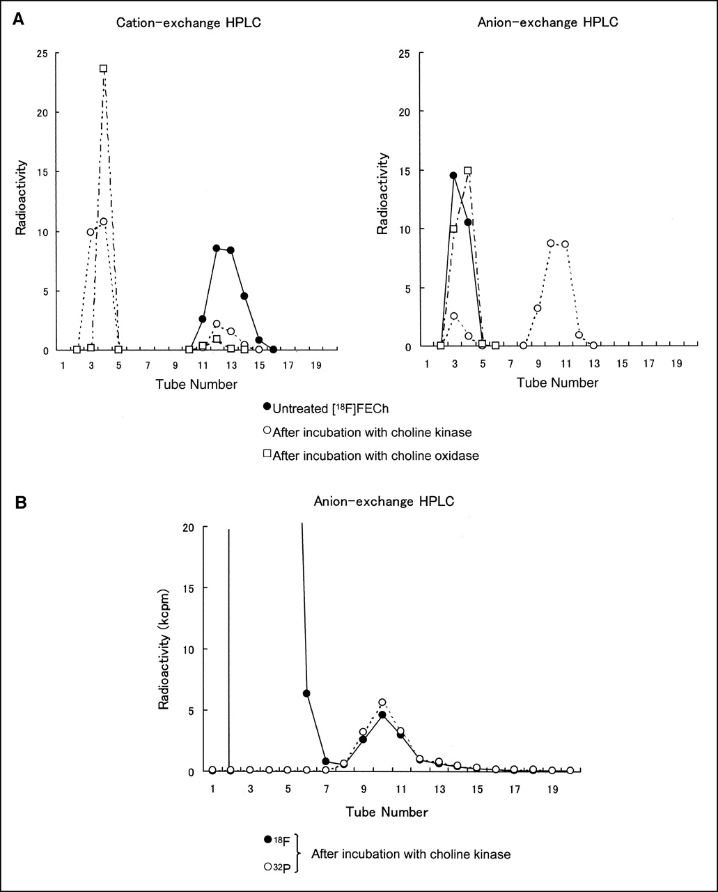
(A) HPLC after incubation of 18F-FECh with choline kinase and with choline oxidase. No-carrier-added 18F-FECh chloride was incubated with yeast choline kinase and 15 μmol ATP or with bacterial choline oxidase. From incubated specimen, methanol-water–soluble component was separated and fractionated on cation- and anion-exchange HPLC. 18F radioactivity was measured in each fraction. (B) HPLC after incubation of 18F-FECh with choline kinase and γ-32P-ATP. No-carrier-added 18F-FECh chloride (approximately 0.005 μmol) was incubated with yeast choline kinase and 0.01 μmol γ-32P-ATP. From incubated specimen, methanol-water–soluble component was separated and fractionated on anion-exchange HPLC. 18F and 32P radioactivities were measured in each fraction. kcpm = kilocounts per minute.
Formation of Fluoroethylbetaine by Choline Oxidase.
A similar experiment was conducted using choline oxidase, in which choline kinase was replaced by choline oxidase, and ATP was omitted. When HPLC was performed, most radioactivity migrated fast on the cation-exchange HPLC and the anion-exchange HPLC (Fig. 3A). These findings presented a great contrast to the result of the choline kinase experiment. This experiment seemed to indicate that 18F-FECh was converted into 18F-fluoroethylbetaine by choline oxidase.
Formation of 32P-Phosphoryl-18F-FECh by Choline Kinase and γ-32P-ATP.
In the above experiment, it was shown that 18F-FECh was converted into a new compound by the reaction with choline kinase and ATP and that the new compound could be considered tentatively as phosphoryl-18F-FECh. In the following experiment, the chemical property of this new compound was studied more precisely. No-carrier-added 18F-FECh (containing approximately 0.005 μmol FECh) was incubated with yeast choline kinase, γ-32P-ATP (0.01 μmol), MgCl2, and dithiothreitol. (The radioactivities of 18F-FECh and γ-32P-ATP added were 12.3 and 0.088 MBq, respectively.) After the reaction was stopped, the methanol-water layer was concentrated and analyzed on the HPLC using anion-exchange resin. Figure 3B shows the result of the HPLC. When the radioactivity of 18F was measured, 2 components were found: a large component that migrated fast and a small component that migrated slowly. When the radioactivity of 32P was measured, no radioactivity was in the fast component but there was distinct radioactivity in the slow component, and the ratio of 32P to 18F in the slow component was even in every fraction. It was evident from this observation that 32P and 18F were tagged by the same molecule. In other words, the new compound produced by the reaction of 18F-FECh with choline kinase and γ-32P-ATP was undoubtedly 32P-phosphoryl-18F-FECh.
Formation of Phosphoryl-18F-FECh in Tumor Cells.
No-carrier-added 18F-FECh chloride was incubated with Ehrlich ascites tumor cells (approximately 106 cells/mL) at 37°C for 30 min. After incubation, the cells were washed by centrifugation, and the methanol-water–soluble fraction was obtained. This fraction was divided into halves, and their radioactivities were analyzed on HPLC: one half on cation-exchange HPLC and the other half on anion-exchange HPLC. Figure 4 shows the result: The cation-exchange HPLC showed that approximately 17.1% of the original 18F-FECh (slowly migrating) was converted into a fast-migrating component (or components), ascribable to either phosphoryl-18F-FECh or 18F-fluoroethylbetaine. The anion-exchange HPLC showed that approximately 17.5% of the original 18F-FECh (fast migrating) was converted into a slowly migrating component, ascribable only to phosphoryl-18F-FECh. This observation indicated that the fast-migrating component in the cation-exchange HPLC was totally ascribable to phosphoryl-18F-FECh. It also indicated that 18F-FECh was converted into phosphoryl-18F-FECh, but not into 18F-fluoroethylbetaine, in this tumor type.

HPLC of methanol-water layer after incubation of 18F-FECh with Ehrlich ascites tumor cells. No-carrier-added 18F-FECh chloride was incubated with Ehrlich ascites tumor cells (approximately 106 cells/mL) in glucose-fortified Hanks’ solution for 30 min. Methanol-water–soluble component was separated from incubated cells and divided into halves. One half was analyzed on HPLC using cation-exchange resin and other half was analyzed on HPLC using anion-exchange resin. 18F radioactivity was measured in every fraction.
Formation of 18F-FECh-Derived Phospholipids in Tumor Cells.
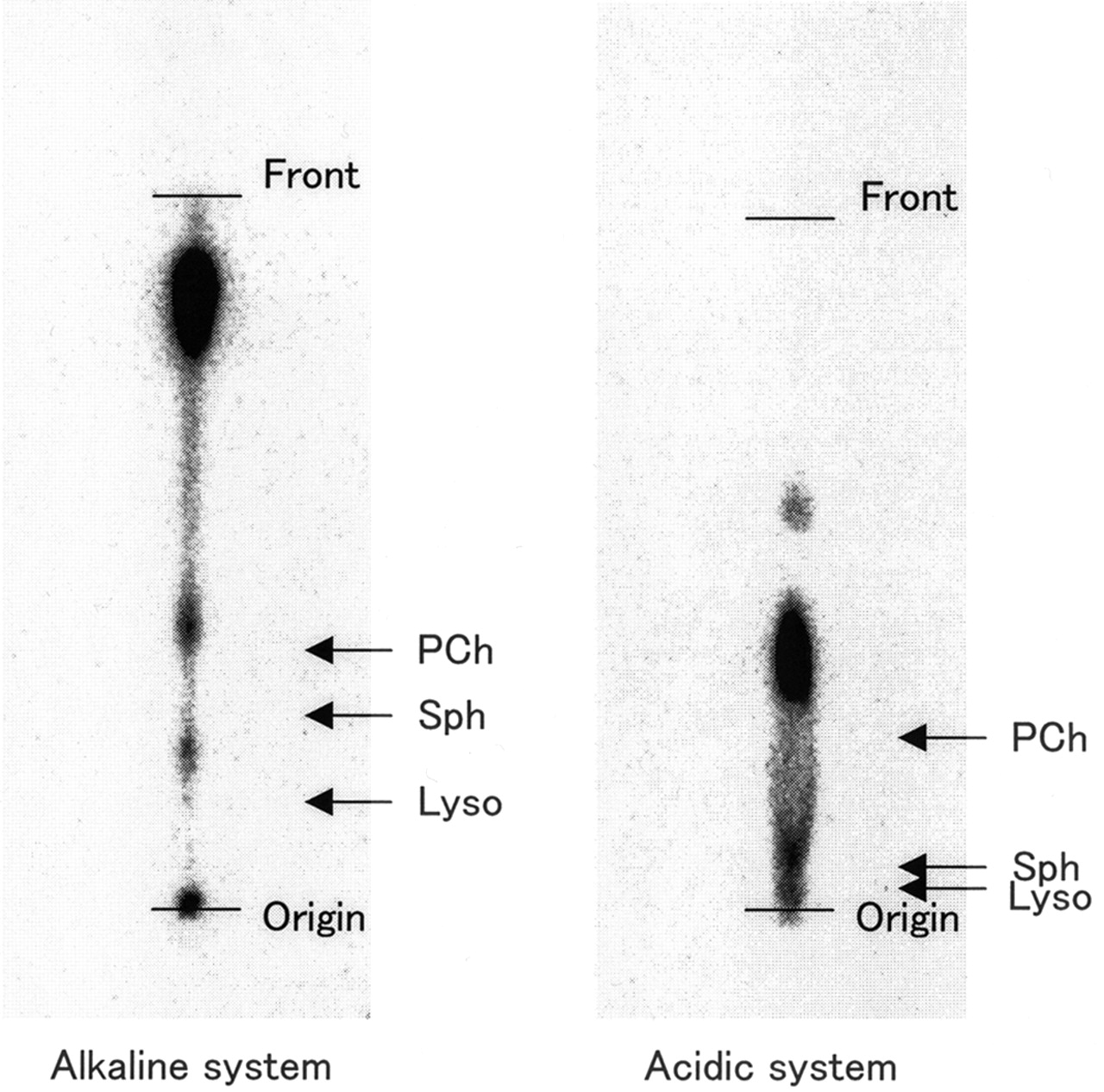
Αfter the above experiment in which the methanol-water–soluble fraction of the tumor cell extract was analyzed, the chloroform-soluble fraction of the tumor cell extract, also containing radioactivity, was analyzed. We thought that all radioactivity in this fraction was from choline-linked phospholipids. No-carrier-added 18F-FECh chloride was incubated with Ehrlich ascites tumor cells (approximately 106 cells/mL) at 37°C for 30 min. After incubation, the cells were washed, and the chloroform-soluble fraction was obtained. The sample was chromatographed then on silica gel TLC plates with 2 solvent systems (alkaline and acidic solvent systems), and the radioactivity distribution on the plate was measured using an imaging-plate-scanner system. Regardless of the solvent system used, the radioactivity was always localized in ≥3 spots, with most concentrated in 1 spot (Fig. 5). Natural choline-containing phospholipids (phosphatidylcholine, sphingomyelin, and lysophosphatidylcholine) migrated to locations very different from the locations of the radioactive spots (Fig. 5, arrows). Nevertheless, we assumed that our radioactive spots corresponded to the natural phospholipids but migrated faster than the natural phospholipids. This assumption was well justified because it is known that the long alkyl group is more hydrophobic than the shorter ones and the fluorinated alkyl group is more hydrophobic than the nonfluorinated ones (13,14). In this context, it was also assumed that the major radioactive spot of our specimen was phosphatidyl-18F-FECh.

TLC of chloroform layer after incubation of 18F-FECh with Ehrlich ascites tumor cells (autoradiography). No-carrier-added 18F-FECh chloride was incubated with Ehrlich ascites tumor cells (approximately 106 cells/mL) in glucose-fortified Hanks’ solution for 30 min. Chloroform-soluble fraction of cell extract was concentrated and developed on TLC silica gel plates. Solvent was as follows: for alkaline system, chloroform, methanol, and 28% ammonia (65:35:5); for acidic system, benzene, pyridine, and formic acid (50:40:10). Autoradiography of TLC plates was performed using imaging plate detector system. Authentic choline-containing phospholipids were developed in same way as above and are indicated by arrows. PCh = phosphatidylcholine; Sph = sphingomyelin; Lyso = lysophosphatidylcholine.
Uptake and Metabolism of 18F-FECh in Tumor Cells.
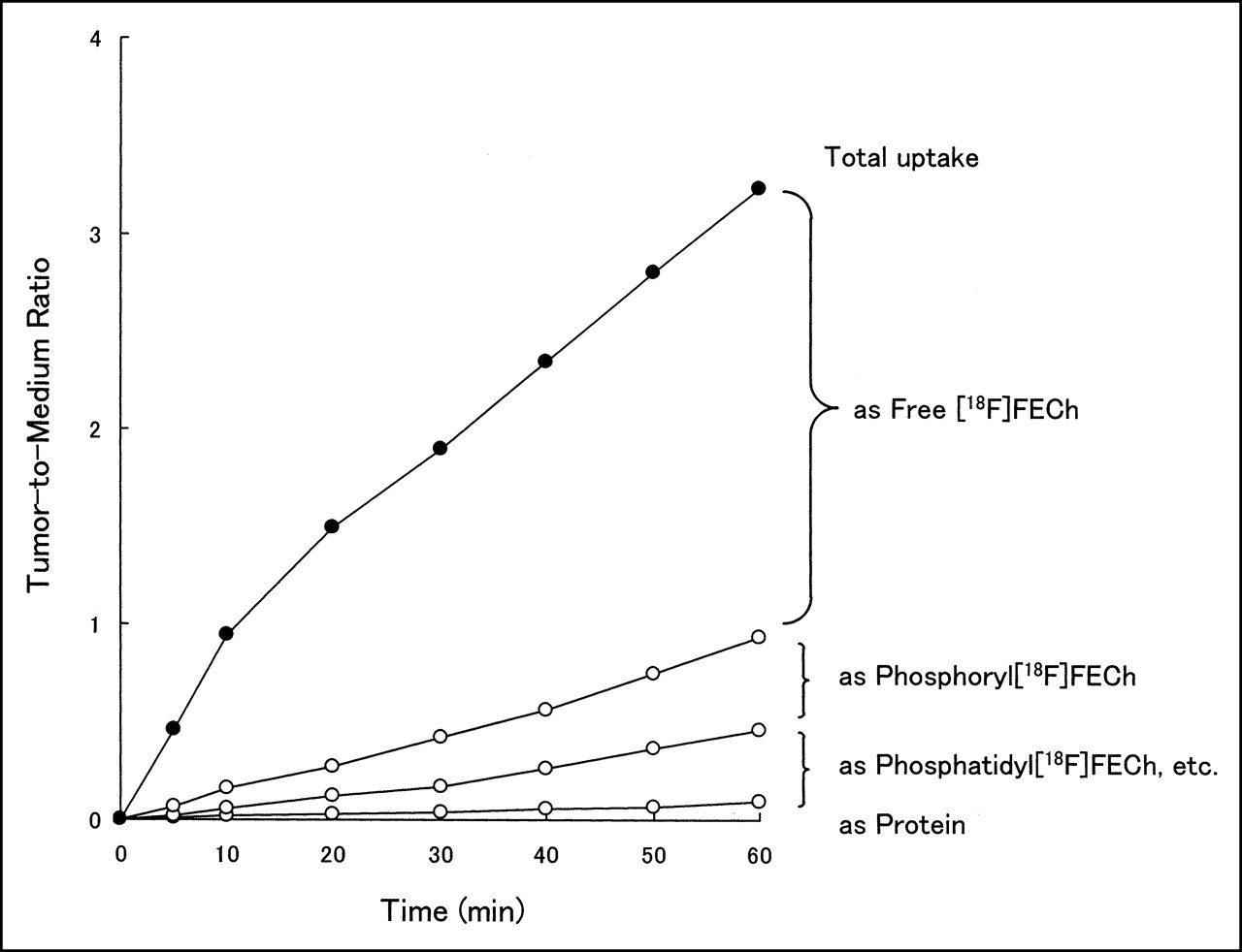
No-carrier-added 18F-FECh chloride was incubated with Ehrlich ascites tumor cells (approximately 106 cells/mL) at 37°C for various times up to 60 min. The total uptake of radioactivity in the cells was measured after centrifugation of the cells, and the cell-to-medium ratio was calculated from the data of the radioactivity of the total cells, the radioactivity of the total medium, and the hematocrit value of the incubation mixture. The washed cells were treated then with methanol and chloroform. After the methanol-water layer was diluted with water and passed through anion-exchange cartridges, the radioactivities in the effluent (free 18F-FECh) and within the cartridge (phosphoryl-18F-FECh) were measured. The radioactivities of the chloroform layer (phospholipids) and the precipitate in the interface (proteins) were also measured. The overall results showed that 18F-FECh moved across the cell membranes into the intracellular space against a concentration gradient (active transport); then, inside the cells, free 18F-FECh was converted gradually into phosphoryl-18F-FECh and finally integrated into phospholipids, primarily into phosphatidyl-18F-FECh. In addition, a small portion of radioactivity was incorporated into proteins (Fig. 6).

Time course of uptake and metabolism of 18F-FECh in Ehrlich ascites tumor cells. No-carrier-added 18F-FECh chloride was incubated with Ehrlich ascites tumor cells (approximately 106 cells/mL) in glucose-fortified Hanks’ solution for various periods up to 60 min. Total 18F uptake was measured after washing cells by centrifugation, and cell-to-medium ratio was calculated after hematocrit determination. After treatment of cells with methanol and chloroform, radioactivities in free 18F-FECh, phosphoryl-18F-FECh, 18F-labeled phospholipids (phosphatidyl-18F-FECh and so forth), and proteins were measured.
Clinical Study
18F-FECh Distribution in a Prostate Cancer Patient.
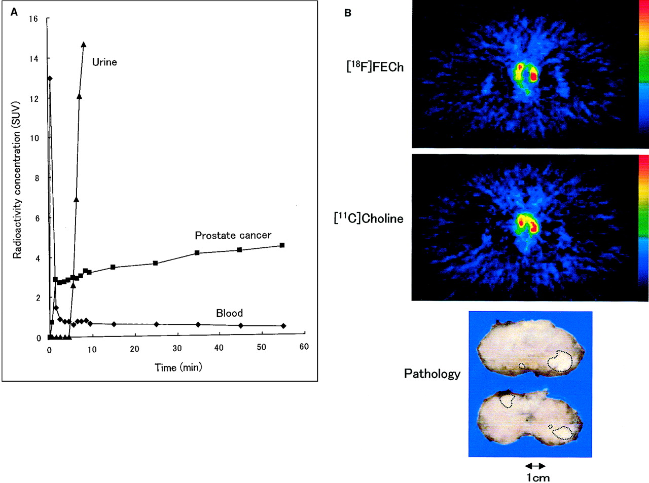
A dynamic PET scan was obtained on a patient with untreated prostate cancer after intravenous injection of 18F-FECh chloride without bladder irrigation. The distribution of radioactivity with time in various organs, including aortic blood, prostate cancer, urine in the bladder (at the orifice of ureter), was determined (Fig. 7A). The blood clearance was rapid, and the blood level became close to minimal in 1 min. The highest radioactivity was found in the kidneys and liver, and no radioactivity was found in the bones (neither in the epiphysis nor in the bone morrow). A large amount of radioactivity appeared in urine after a time lag of 5 min. The prostate cancer showed the highest radioactivity among all intrapelvic organs except for urine in the bladder. The uptake of radioactivity in prostate cancer was rapid and showed an SUV of 2.87 at 1.5 min; then it increased slowly, giving an SUV of 4.43 at 55 min after injection.

(A) Time course of radioactivity concentration in blood, prostate cancer, and urine after intravenous injection of 18F-FECh to untreated prostate cancer patient. SUV was calculated from PET data. (B) PET images of prostate cancer with 18F-FECh and 11C-choline and pathologic specimen of resected prostate (patient 5). 18F-FECh and 11C-choline images were taken at 60 and 20 min after injection, respectively. Red represents SUV of ≥4.0. Under diagnosis of stage B2, total prostatectomy and bilateral pelvic lymph node dissection were performed. Pathologic examination indicated adenocarcinoma of prostate in both lobes (Gleason score, 7) and no metastasis in pelvic lymph nodes (pathologic stage, pT2b, pN0). Largest tumor mass, 1.2 cm in diameter, was located in peripheral zone of left lobe.
18F-FECh PET and 11C-Choline PET in Prostate Cancer Patients.
Static PET scans were obtained on 16 patients with untreated primary prostate cancer. The radioactivity in urine was eliminated by continuous bladder irrigation. The radioactivity of prostate cancer expressed by SUV was essentially unchanged between 30 and 60 min after injection. Radioactivity appeared sometimes in the intestinal fluid, for which the location and intensity changed with time. This artifact was easily discriminated from the true uptake of prostate cancer by comparison of the 30- and 60-min images (our computer program facilitated this discrimination). Table 1 shows the data of the patients with untreated prostate cancer: number of positive (malignant) samples of biopsy from all samples examined (transrectal sonographically guided needle biopsy of the prostate), grade of tumor cell differentiation in the biopsy samples (poorly, moderately, or well differentiated), level of prostate-specific antigen (PSA) in the blood, and clinical stage (15) estimated from the findings of biopsy, PET, and other modalities. Table 1 also shows the SUVs in the most radioactive area in the region of the prostate, determined by PET with 18F-FECh and with 11C-choline, where the 18F-FECh image was taken at 30 and 60 min after injection and the 11C-choline image was taken at 5 and 20 min after injection, respectively. Of all 16 patients examined, 1 patient (patient 5, Fig. 7) underwent total prostatectomy immediately after the PET study, and all others were treated thereafter by hormonal therapy as an immediate measure.
Patients with Primary Prostate Cancer
With both 18F-FECh and 11C-choline, the prostate cancer was always visualized as high-uptake areas surrounded by low-uptake areas of normal prostate, where the high-uptake and low-uptake areas of the PET image corresponded well to the malignant and normal findings of the biopsy examination. With 18F-FECh, the tumor SUV (within an area of 8 × 8 mm in real size) ranged from 1.97 to 6.47 (mean ± SD, 3.84 ± 1.25) at 30 min and from 1.86 to 7.50 (4.02 ± 1.46) at 60 min after injection, respectively. With 11C-choline, the tumor SUV ranged from 1.66 to 7.22 (4.03 ± 1.38) at 5 min and from 1.86 to 7.58 (4.50 ± 1.56) at 20 min after injection, respectively. With 18F-FECh and 11C-choline, the SUVs stayed unchanged during the 2 scan times. In addition, in the area of the cancer, the SUV of 18F-FECh was almost the same as the SUV of 11C-choline.
In general, 18F-FECh PET gave slightly better images of prostate cancer than 11C-choline PET in terms of spatial resolution. This observation is explainable by the shorter positron range of 18F.
DISCUSSION
We reported, in a preliminary form, the synthesis of 18F-FECh (2-fluoroethyl-dimethyl-2-oxyethylammonium) (12), in which we used Kryptofix 2.2.2. (Merck-Schuchardt, Hohenbrunn, Germany) as the synthetic catalyst. In this study, we used TBA bicarbonate instead of Kryptofix 2.2.2. because we were concerned that Kryptofix 2.2.2. might deteriorate the purity of 18F-FECh. Our apprehensions were based on the following facts: First, because the behavior of Kryptofix 2.2.2. in preparative HPLC was similar to that of 18F-FECh (Kryptofix 2.2.2. migrated just behind 18F-FECh), it might spill over into the 18F-FECh peak (TBA migrated far behind the 18F-FECh peak). Second, because Kryptofix 2.2.2. contains 2 tertiary amine moieties, they might react with 2-18F-fluoroethyl tosylate to produce undesirable by-products. Using TBA bicarbonate, these apprehensionswere eliminated, and the radiochemical and chemical purities increased markedly, but the radiochemical yield and the time of synthesis did not change. We constructed an automated apparatus for the synthesis of 18F-FECh chloride, according to the TBA method, and examined its performance >50 times: The reliability of the apparatus was excellent (no failure), and the radiochemical yield always stayed within the range of 35%–45%, with the decay corrected.
In the in vitro experiment, we confirmed that 18F-FECh was incorporated into tumor cells by active transport (against the concentration gradient); then phosphorylated inside the cells, yielding phosphoryl-18F-FECh; and finally integrated into phospholipids, probably primarily into phosphatidyl-18F-FECh. These mechanisms constitute a kind of chemical trap and seem to explain why 18F-FECh was incorporated into prostate cancer so markedly and why the SUV in prostate cancer remained constant for a long time. When the SUV of 18F-FECh in prostate cancer of a patient was compared with the SUV of 11C-choline in prostate cancer of the same patient, these values were always close to each other. This fact seemed to indicate that the mechanisms of the membrane transport of 18F-FECh and 11C-choline are the same, and the mechanisms of the phosphorylation of 18F-FECh and 11C-choline are also the same; and the biologic activities of these mechanisms for these 2 substrates are almost the same, whereas the uptake mechanism of 11C-choline in tumors has been well discussed by us (5–7,9) and by others (16,17).
The advantages of 18F-FECh over 11C-choline were as follows. First, because of the longer half-life of 18F, 18F was more convenient for long-time storage and long-distance transportation. Second, because of the shorter positron range of 18F, 18F gave a slightly higher quality of image with higher spatial resolution.
The disadvantage of 18F-FECh was the rapid excretion of radioactivity into urine (in contrast to 11C-choline), and it was necessary to irrigate the bladder continuously using a urinary catheter to eliminate the bladder radioactivity. However, this procedure was very uncomfortable for urology patients.
DeGrado et al. (18) synthesized 18F-fluoromethylcholine and observed its biodistribution in mice with prostate cancer xenografts. They reported a high uptake of this compound in tumors, and a high radioactivity in urine that was 10 times higher than that of 11C-choline at 30 min after injection.
Recently, DeGrado et al. (19) reported on their success in obtaining clear PET images of prostate cancer in patients in whom 18F-fluoromethylcholine was used instead of 18F-FECh, the bladder irrigation was avoided, and the scanning was conducted at 3–5 min after injection (before the emergence of radioactivity in urine). We also obtained a clear PET image of prostate cancer in 1 patient (Fig. 7A) using 18F-FECh, without bladder irrigation, and conducting the scanning at 2–5 min after injection. We did not adopt this protocol in the rest of this study because we wanted to examine the whole area of pelvis in the patients (our PET machine covers only 6.5 cm longitudinally at 1 bed position).
CONCLUSION
It is established that 11C-choline PET is very effective in detecting various cancers, including prostate cancer. We developed a method to synthesize 18F-FECh as a substitute for 11C-choline and constructed an automated apparatus for the synthesis. In addition, we studied the tumor uptake of 18F-FECh in Ehrlich tumor cells in vitro and performed 18F-FECh PET on 16 patients with untreated primary prostate cancer. Our method of synthesis was easy and reliable, and the performance of our automated apparatus was excellent. In the in vitro experiment, 18F-FECh was incorporated into the tumor cells by active transport, then phosphorylated within the cells, and finally integrated into phospholipids, constituting a chemical trap mechanism in the tumor cells. In the clinical study, 18F-FECh PET visualized prostate cancer of the patients at the same uptake rate (SUV) as that observed by 11C-choline PET. However, 18F-FECh PET was slightly superior to 11C-choline PET in the sharpness of the image. The only disadvantage of 18F-FECh PET compared with 11C-choline PET was the need to introduce a urinary catheter into the bladder for continuous withdrawal of urine during the time of PET scanning.
Acknowledgments
This work was supported in part by the Ministry of Education, Culture, Sports, Science and Technology of Japan and the Japanese Smoking Research Foundation.
Footnotes
Received Mar. 1, 2001; revision accepted Jun. 14, 2001.
For correspondence or reprints contact: Toshihiko Hara, MD, PhD, Department of Radiology, International Medical Center of Japan, 1-21-1 Toyama, Shinjuku-ku, Tokyo 162, Japan.
E-mail: ahara{at}kt.rim.or.jp
REFERENCES
In this issue


Jump to section
Related Articles
Cited By...
- Molecular Imaging of Prostate Cancer: Tapping into the Opportunities
- Combined PET/MRI Improves Diagnostic Accuracy in Patients with Prostate Cancer: A Prospective Diagnostic Trial
- Reduced 64Cu Uptake and Tumor Growth Inhibition by Knockdown of Human Copper Transporter 1 in Xenograft Mouse Model of Prostate Cancer
- Detection of Recurrent Prostate Cancer After Radical Prostatectomy: Comparison of 11C-Choline PET/CT with Pelvic Multiparametric MR Imaging with Endorectal Coil
- Choline phosphorylation and regulation of transcription of choline kinase {alpha} in hypoxia
- Novel Tracers and Their Development for the Imaging of Metastatic Prostate Cancer
- Gene Expression Patterns and Tumor Uptake of 18F-FDG, 18F-FLT, and 18F-FEC in PET/MRI of an Orthotopic Mouse Xenotransplantation Model of Pancreatic Cancer
- Tumor Cell Metabolism Imaging
- Choline Kinase Down-regulation Increases the Effect of 5-Fluorouracil in Breast Cancer Cells
- Initial Experience with the Radiotracer Anti-1-Amino-3-18F-Fluorocyclobutane-1-Carboxylic Acid with PET/CT in Prostate Carcinoma
- Imaging Prostate Cancer with 11C-Choline PET/CT
- Value of 11C-Choline PET and Contrast-Enhanced CT for Staging of Bladder Cancer: Correlation with Histopathologic Findings
- Localization of Primary Prostate Cancer with Dual-Phase 18F-Fluorocholine PET
- Phosphorylation of the Yeast Choline Kinase by Protein Kinase C: IDENTIFICATION OF Ser25 AND Ser30 AS MAJOR SITES OF PHOSPHORYLATION
- microPET and Autoradiographic Imaging of GRP Receptor Expression with 64Cu-DOTA-[Lys3]Bombesin in Human Prostate Adenocarcinoma Xenografts
- PET Scan Detects Prostate Cancer in a Patient with Hodgkins Lymphoma
- PET Imaging of Prostate Cancer with 11C-Acetate
- 11C-Acetate PET Imaging of Prostate Cancer: Detection of Recurrent Disease at PSA Relapse
- Phosphorylation of Saccharomyces cerevisiae Choline Kinase on Ser30 and Ser85 by Protein Kinase A Regulates Phosphatidylcholine Synthesis by the CDP-choline Pathway
- PET for Prostate Cancer Imaging: Still a Quandary or the Ultimate Solution?