

FIGURE 5.
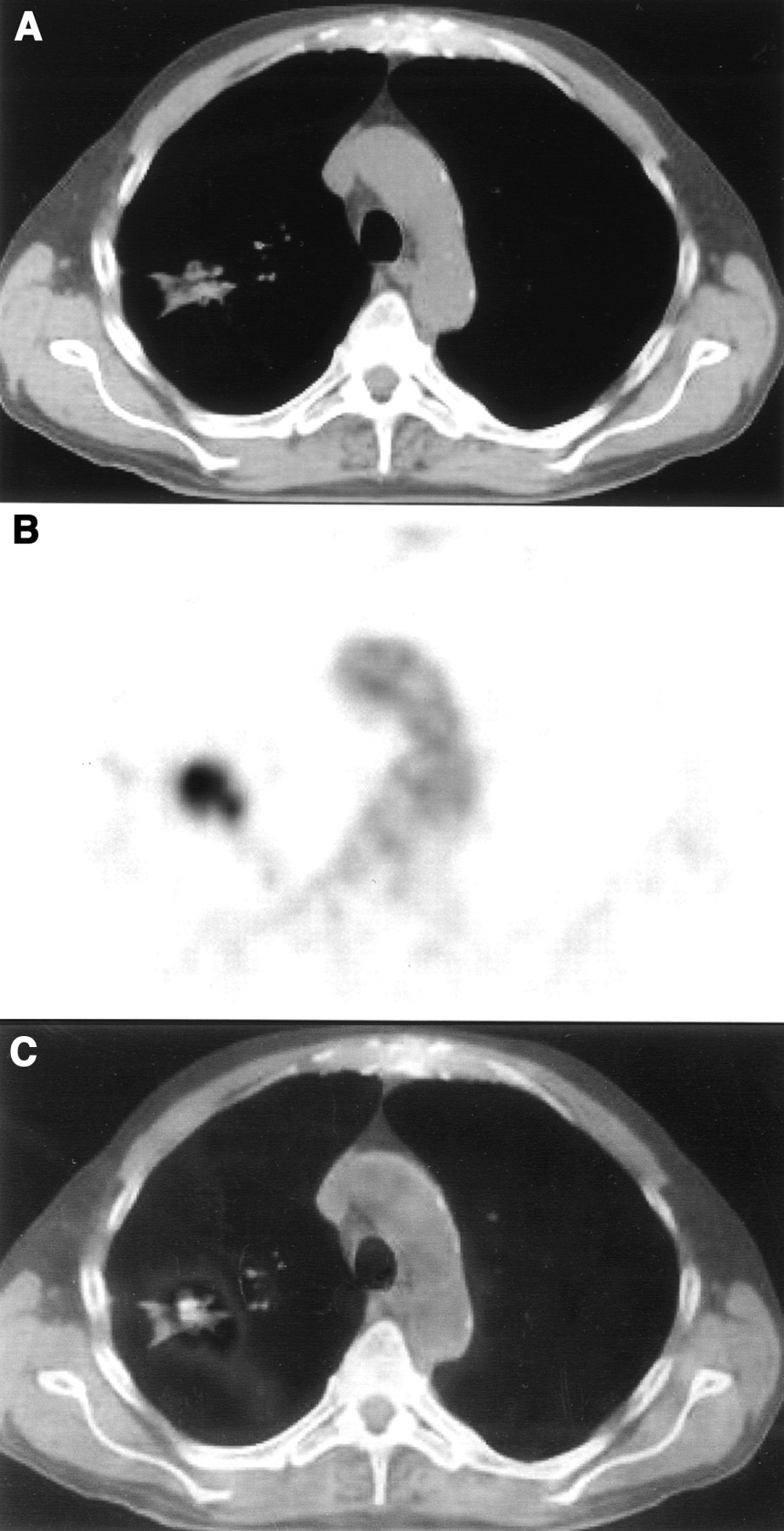
FIGURE 5.
Patient with solitary lung lesion caused by NSCLC in upper peripheral part of right lung. Coregistration of CT (A) and PET (B) images is visually adequate, but measured incongruence was 1.8 mm. This is due to different shapes of lesions, with more star-like appearance on CT and roundly shaped lesion on PET. (C) Final coregistered PET/CT image.
In this issue


Related Articles
Cited By...
- Improving the Spatial Alignment in PET/CT Using Amplitude-Based Respiration-Gated PET and Patient-Specific Breathing-Instructed CT
- Value of 4-Dimensional 18F-FDG PET/CT in the Classification of Pulmonary Lesions
- Latest Advances in Molecular Imaging Instrumentation
- Use of H215O-PET and DCE-MRI to Measure Tumor Blood Flow
- Postacquisition Detection of Tumor Motion in the Lung and Upper Abdomen Using List-Mode PET Data: A Feasibility Study
- Imaging and Oncologic Drug Development
- Automated 3-Dimensional Elastic Registration of Whole-Body PET and CT from Separate or Combined Scanners
- Attenuation Correction of PET Images with Respiration-Averaged CT Images in PET/CT
- The CT Motion Quantitation of Lung Lesions and Its Impact on PET-Measured SUVs
- Automated 3-Dimensional Registration of Stand-Alone 18F-FDG Whole-Body PET with CT