


Abstract
Early and accurate diagnosis of osteomyelitis remains a clinical problem. Acute osteomyelitis often occurs in infants and most often is located in the long bones. Radiologic images show changes only in advanced stages of disease. Scintigraphic imaging with 99mTc-methylene diphosphonate (MDP), or bone scanning, is much more sensitive in detecting acute osteomyelitis but lacks specificity. We evaluated the performance of 99mTc-interleukin-8 (IL-8) in an experimental model of acute osteomyelitis. Methods: Acute pyogenic osteomyelitis was induced in 10 rabbits by inserting sodium morrhuate and Staphylococcus aureus into the medullary cavity of the right femur. The cavity was closed with liquid cement. A sham operation was performed on the left femur. Routine radiographs were obtained just before scintigraphy. Ten days after surgery, the rabbits were divided into 2 groups of 5 animals, received an injection of either 18.5 MBq 111In-granulocytes or 18.5 MBq 67Ga-citrate, and were imaged both 24 h after injection and 48 h after injection. On day 12, the rabbits received either 18.5 MBq 99mTc-MDP or 18.5 MBq 99mTc-IL-8, and serial images were acquired at 0, 1, 2, 4, 8, 12, and 24 h after injection. Uptake in the infected femur was determined by drawing regions of interest. Ratios of infected femur (target) to sham-operated femur (background) (T/Bs) were calculated. After the final images were obtained, the rabbits were killed and the right femur was dissected and analyzed for microbiologic and histopathologic evidence of osteomyelitis. Results: Acute osteomyelitis developed in 8 of 10 rabbits. All imaging agents correctly detected the acute osteomyelitis in these animals. The extent of infection was optimally visualized with 67Ga-citrate and delayed bone scanning, whereas diaphyseal photopenia was noted with both 99mTc-IL-8 and 111In-granulocytes. In 1 rabbit with osteomyelitis, imaging results were falsely negative with 111In-granulocytes and falsely positive with 99mTc-MDP. Quantitative analysis of the images revealed that the uptake in the infected region was highest with 67Ga-citrate (4.9 ± 0.8 percentage injected dose [%ID]) and 99mTc-MDP (4.7 ± 0.7 %ID), whereas the uptake in the infected area was significantly lower with 99mTc-IL-8 (2.2 ± 0.2 %ID) and 111In-granulocytes (0.8 ± 0.2 %ID) (P < 0.0042). In contrast, the T/Bs were significantly higher for 99mTc-IL-8 (T/B, 6.2 ± 0.3 at 4 h after injection) than for 67Ga-citrate, 99mTc-MDP, and 111In-granulocytes, which had ratios of 1.5 ± 0.4, 1.9 ± 0.2, and 1.4 ± 0.1, respectively (P < 0.0001). Radiography correctly revealed acute osteomyelitis in only 2 of 8 rabbits. Conclusion: In this rabbit model of osteomyelitis, 99mTc-IL-8 clearly revealed the osteomyelitic lesion. Although the absolute uptake in the osteomyelitic area was significantly lower than that obtained with 99mTc-MDP and 67Ga-citrate, the T/Bs were significantly higher for 99mTc-IL-8 because of fast background clearance. The ease of preparation, good image quality, and lower radiation burden suggest that 99mTc-IL-8 may be a suitable imaging agent for the scintigraphic evaluation of acute osteomyelitis.
In cases of delayed therapy, acute osteomyelitis can, within several days, cause severe malformations of the bone that can become disabling and substantially affect the quality of life (1). Acute osteomyelitis usually is diagnosed on the basis of imaging, laboratory tests, and clinical examinations (2–5). In nuclear medicine, 99mTc-methylene diphosphonate (MDP) and 67Ga-citrate are sensitive agents for the detection of osteomyelitis. Their specificity, however, is low because both agents accumulate in any area with increased bone turnover (6). Radiolabeled white blood cells (7,8) as imaging agents have a much higher specificity for scintigraphic imaging of osteomyelitis. The preparation of radiolabeled autologous white blood cells, however, is laborious and time consuming and carries a small but definite risk of personal contamination by the patients’ blood and inadvertent cross-contamination between patients (9,10).
New radiopharmaceuticals, such as radiolabeled monoclonal antigranulocyte antibody preparations, have been proposed for easy and fast imaging of infection (11,12). These new agents displayed high sensitivity and specificity in patients with acute osteomyelitis (13–15), presumably because they act through cell-specific labeling of surface antigens as present on granulocytes.
Granulocytes are known to express interleukin-8 (IL-8) receptors abundantly (16–19). Therefore, the proinflammatory chemotactic cytokine IL-8 may be an interesting alternative to the current methods for imaging acute osteomyelitis.
Recently, we introduced IL-8 as a new scintigraphic imaging agent. In a rabbit model with Escherichia coli soft-tissue infection, 99mTc-hydrazinonicotinamide-IL-8 allowed rapid visualization of the infectious foci as early as 1 h after injection, with high and rapid accretion of the radiolabel in the abscess (20). This study was performed primarily to gain our first preclinical experience with the use of this new 99mTc-IL-8 tracer to detect experimentally induced osteomyelitis. Therefore, we evaluated 99mTc-IL-8 as an imaging agent in a rabbit model of acute osteomyelitis. The results were compared with those obtained using the conventional and well-established agents 99mTc-MDP, 67Ga-citrate, and 111In-granulocytes.
MATERIALS AND METHODS
Animal Osteomyelitis Model
This study was performed in accordance with the guidelines of the local animal welfare committee. Adult female New Zealand White rabbits (weight range, 2.5–3.0 kg) were obtained from the central animal laboratory, University of Nijmegen (Nijmegen, The Netherlands), caged individually, and fed regular rabbit diet and water ad libitum. In 10 rabbits, acute osteomyelitis was induced as described previously, with minor modifications (21–23). The rabbits were anesthetized with a mixture of halothane, nitrous oxide, and oxygen and placed prone on the operation table. Both hind legs were shaved, disinfected with a 2% tincture of iodine, and isolated by sterile drapes. The trochanter tertius was exposed bilaterally, and the cortex was penetrated gently using a hand drill. A small syringe with a 2-mm-long silicone tube (outer diameter, 3.0 mm) was inserted into the femoral canal, and 0.5 mL 5% sodium morrhuate (QUAD Pharmaceuticals Inc., Indianapolis, IN) was inoculated in the canal. Morrhuate, a complex of fatty acids rich in arachidonic acid (24,25), was used to induce a local irritation. Subsequently, 0.5 mL of 5 × 10⁶ colony-forming units of Staphylococcus aureus (ATCC 25923; American Type Culture Collection, Manassas, VA) was inoculated. For the sham procedure on the right femur, the canal was washed with 1 mL saline and left without a local irritation/infection procedure. Finally, both holes in the trochanter were sealed with a small amount of liquid carboxylate cement (Durelon; ESPE Dental AG, Seefeld, Germany). After polymerization of the cement, the wounds at both sides were cleaned with sterile saline solution and closed. The animals were examined regularly with special attention to wound healing, body temperature, and body weight.
Radiopharmaceuticals
99mTc-IL-8.
The 99mTc-labeled IL-8 was prepared as described previously (20), with minor modifications. Briefly, to a 1.5-mL vial containing 6 μg IL-8 was added 0.4 mL tricine solution (N-[Tris(hydroxymethyl)methyl]glycine) (Fluka, Buchs, Switzerland; 100 mg/mL in 25 mmol/L succinate buffer, pH 5.0) and 0.1 mL isonicotinic acid solution (Sigma, St. Louis, MO; 20 mg/mL in 25 mmol/L succinate buffer, pH 5.0) (26). After the reaction mixture had been purged with a gentle stream of nitrogen, 25 μL SnSO4 solution (1.0 mg/mL in 0.1N HCl) and 350 MBq 99mTcO4− were added. After having been heated to 70°C for 30 min, the reaction mixture was cooled to room temperature and the radiochemical purity was determined by instant thin-layer chromatography (ITLC) on ITLC-SG strips (Gelman Laboratories, Ann Arbor, MI) with 0.1 mol/L citrate, pH 6.0, as the mobile phase.
The 99mTc-IL-8 was purified on a Sephadex G-25 column (PD-10; Pharmacia, Uppsala, Sweden) eluted with 0.5% bovine serum albumin in phosphate-buffered saline. The labeling efficiency of the 99mTc-IL-8 preparation exceeded 90%. After Sephadex G-25 purification, the radiochemical purity of the radiopharmaceutical exceeded 98% as determined by ITLC. The specific activity of the purified preparation was 50 MBq/μg (425 GBq/μmol).
111In-Granulocytes.
Carotid artery cannulation was performed on 1 anesthetized donor rabbit. A total of 100 mL blood was carefully drawn into acid citrate dextrose tubes (containing 7 mL acid citrate dextrose per 35 mL blood). The total leukocyte count of the donor rabbits was 6.8 × 10⁹ cells/L, with approximately 50% granulocytes. The granulocytes were purified according to the method described by Lillevang et al. (27), with minor modifications (28). Briefly, the blood was mixed with 0.1 volume of 6% dextran (Dextran 500; Pharmacia) solution in 0.9% NaCl and allowed to settle for 1 h at room temperature. The leukocyte-rich supernatant was layered carefully on 0.3 volume of Nycoprep density medium (Nycomed, Oslo, Norway; 14.1% Nycodenz [Nycomed], 0.3% NaCl, 5 mmol/L tricine/NaOH, pH 7.2, 1.077 g/mL density, 265 mOsm) and centrifuged for 15 min at 600g. The plasma above the mononuclear cells, the mononuclear band, and the density medium above the granulocyte pellet were carefully removed. The pellet was washed with 5 mL Hanks’ balanced salt solution (HBSS) with 10% autologous plasma and centrifuged for 10 min at 50g. The cell pellet was resuspended in 1.5 mL HBSS with 10% rabbit plasma. After this purification procedure, the granulocyte purity was >90%. Subsequently, 185 MBq 111In-oxine were added to the cell suspension. The cells were incubated at room temperature for 30 min and centrifuged for 10 min at 50g. The pellet was resuspended in 5 mL cell-free autologous plasma. Labeling efficiency (cell-associated activity/total activity added) exceeded 80%. The functional integrity of the labeled granulocytes was evaluated by their in vivo performance, including transit through the lungs, as well as uptake in the liver and spleen. A dose of 18 MBq 111In-oxine–labeled granulocytes was administered intravenously to each rabbit.
67Ga-Citrate.
67Ga-citrate (DRN 3103) was purchased from Mallinckrodt, Inc. (St. Louis, MO). A dose of approximately 18 MBq 67Ga-citrate per rabbit was injected intravenously.
99mTc-MDP.
A kit containing MDP and stannous chloride was labeled with 99mTc with a labeling efficiency > 95% as determined by ITLC. A dose of 18 MBq 99mTc-MDP was administered intravenously.
Receptor-Binding Assays
The receptor-binding fraction of the 99mTc-IL-8 preparation was determined in receptor-binding assays essentially as described by Lindmo et al. (29). For receptor-binding assays, Jurkat cells transfected with CXCR1 or CXCR2 were used (30). The cells were cultured at 37°C in a humidified atmosphere of air and CO2 (95:5) in Roswell Park Memorial Institute (RPMI) 1640 medium (GIBCO, Gaithersburg, MD) containing 10% fetal calf serum, 1% glutamine, penicillin and streptomycin, 5 × 10⁻⁵ mol/L β-mercaptoethanol, and 1.5 μg/mL puromycin. Series of serially diluted cell suspension (0.25–4 × 10⁸ cells/mL) were incubated with 10,000 cpm of 99mTc-IL-8 in assay buffer (RPMI 1640, 0.5% bovine serum albumin, and 0.05% NaN3). Duplicates of the lowest cell concentration were incubated in the presence of at least a 100-fold molar excess of unlabeled IL-8 to correct for nonspecific binding. After 30 min of incubation at 37°C, the cells were centrifuged (5 min, 2000g) and the radioactivity in the pellet (total bound radioactivity) was measured in a shielded well scintillation γ-counter (Wizard; Pharmacia). The data were graphically analyzed in a modified Lineweaver–Burk plot: a double-inverse plot of the conventional binding plot (specifically bound fraction vs. cell concentration). The receptor-binding fraction at infinite cell excess was calculated by linear extrapolation to the ordinate. The 99mTc-IL-8 preparation showed a CXCR1 receptor-binding fraction of 55% and a CXCR2 receptor-binding fraction of 45%.
Study Design
The rabbits were randomized into 2 groups: A (n = 5) and B (n = 5). The scintigraphic studies were started 10 d after induction of osteomyelitis. The radiopharmaceuticals were injected in a fixed order. On the first day of the imaging experiment, the rabbits of group A received 18.5 MBq 111In-granulocytes intravenously and the rabbits of group B received 18.5 MBq 67Ga-citrate intravenously. The scintigraphic images were acquired 24 and 48 h after injection of each of the radiotracers. On the third day of the imaging experiment (12 d after induction of osteomyelitis), the rabbits of group A received 18.5 MBq 99mTc-MDP intravenously and the rabbits of group B received 18.5 MBq 99mTc-IL-8 intravenously. Crossover of remaining 67Ga and 111In activity in the 99mTc channel was <5% as determined before and after injection of the 99mTc-labelled compounds. To exclude pharmaceutical interference with the cell-binding capacity of the radiotracer, we did not sedate the rabbits. The rabbits were immobilized in a mold and placed prone on a gamma camera equipped with a medium-energy collimator (Orbiter; Siemens Inc., Hoffman Estates, IL) for the 67Ga and 111In studies and with a parallel-hole, low-energy collimator for the 99mTc-MDP and 99mTc-IL-8 studies. Imaging was performed at 24 and 48 h (for 111In and 67Ga, respectively) after injection. The imaging sessions for 99mTc-MDP and 99mTc-IL-8 were at 3 min (blood-pool image) and 4 h (delayed image) after injection for the bone scans and at 2 min and 1, 2, 4, 8, 12, and 24 h after injection for the serial IL-8 images. Images (300,000 counts per image) were obtained and stored in a 256 × 256 matrix.
The scintigraphic results were analyzed both qualitatively and quantitatively. All scans were evaluated qualitatively without knowledge of the histopathologic outcome. Findings were considered positive if focal uptake of radioactivity at the osteomyelitic site exceeded uptake at the sham-operated site. For quantitative analysis, regions of interest were drawn over the infected right femur and the sham-operated left femur, as well as over the whole body. Additional regions of interest (at 0, 1, 4, 10, and 20 h after injection) over the lungs, liver, and spleen were drawn on the scintigrams of the rabbits that received 111In-granulocytes. Counts were corrected for differences in the number of pixels before ratios and percentages were calculated. The ratios of infected femur (target) to sham-operated femur (background) (T/Bs) were calculated. Residual activity at the osteomyelitic site was determined and measured as fraction of the injected dose (whole body set to t = 0).
Radiologic and Histologic Analyses
The results of the scintigraphic studies were compared with the results of radiologic, microbiologic, and histopathologic examination. Conventional radiographs were obtained after the imaging sessions. While unaware of the results of all other procedures, we evaluated the radiograms with respect to 3 parameters (periosteal thinning, increased radiolucency, and inclusion of air) (31). Immediately after completion of the scintigraphic studies, the rabbits were killed with an overdose of sodium phenobarbital. The right femur of each animal was excised and tissue debris was removed. The femur was halved longitudinally using a high-speed dental drill with a circular metal saw. The bone cement was carefully removed. One bone specimen of each femur was sent for microbiologic examination. For histopathologic examination, the other bone specimens were fixed in 4% buffered formalin and decalcified in 10% ethylenediaminetetraacetic acid. Longitudinal sections were made, mounted on slides, and stained with hematoxylin and eosin. All sections were reviewed microscopically with respect to 3 parameters: purulent inflammation with infiltration of the bone marrow by polymorphonuclear leukocytes, necrotic bone, and bone fistulas. The presence of osteomyelitis was confirmed on the basis of these histopathologic findings.
Statistical Analysis
Mean uptake values are given as percentage injected dose (%ID) ± 1 SEM. For paired data, 1-way ANOVA using InStat software (version 3.00; GraphPad Software, Inc., San Diego, CA) was performed to compare uptake at the osteomyelitic and sham-surgery sites for the different agents. In addition, repeated-measures ANOVA was used to evaluate differences between the images acquired at different times for each agent. The level of significance was set at 0.05.
RESULTS
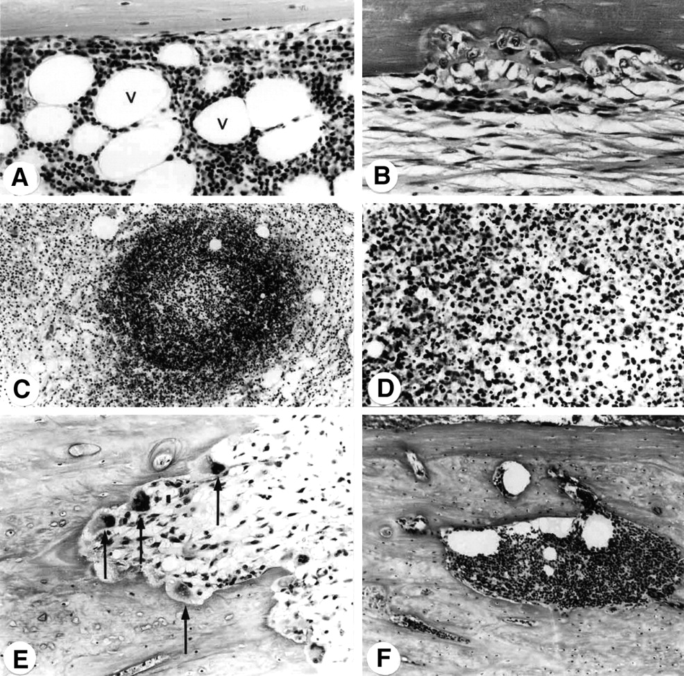
Tables 1 and 2 summarize the observations for groups A and B, respectively. Eight of the 10 rabbits had histopathologic and microbiologic evidence of acute osteomyelitis in the right femur. Macroscopically, in 3 of 8 rabbits the osteomyelitis in the right femur had affected the complete femur, whereas in 5 of 8 rabbits the infection was apparent only in the diaphyseal and distal parts of the femur. The proximal part of the osteomyelitis-negative rabbit (rabbit 3) showed some minimal leukocytic infiltration, which was most likely caused by a local reaction to loosening of the cement. No evidence of infection was found in rabbit 6. The microbiologic studies of the bone specimens were concordant with the histopathologic findings in all infected rabbits and confirmed the presence of S. aureus infection in 8 of 10 rabbits. Histopathologically, the infected medullar tissue was characterized by large accumulations of polymorphonuclear leucocytes (PMNs) and some lymphocytes. Abundant areas of debris and of necrosis were seen. The trabecular bone near the growth plate was, in most cases, necrotic and infiltrated by many PMNs. The cortical bone showed necrosis and infiltration of PMNs, osteoclastic resorption, and a strong periosteal callous formation (Fig. 1). In contrast, radiologic findings were abnormal in only 2 rabbits, showing discrete periosteal thinning and increased radiolucency.

(A) Medullar tissue of control animal containing numerous fat cells (v) and red marrow (×150). (B) Medullar tissue of low-grade infection: fibrous tissue and osteoclastic erosion on endosteal surface of femoral cortex (×150). (C) Large accumulation of PMNs in medullar tissue of heavily infected bone specimen (×50). (D) Enlargement of central part because of PMN accumulation (×250). (E) Erosion channels and osteoclasts (arrows) in cortical infected bone (×100). (F) Eroded areas in cortical bone filled with leukocytes (×80).
Results for Group A
Results for Group B
Quantitative analysis of the scintigrams of 111In-granulocytes showed rapid initial lung transit (uptake = 86 %ID at t0 and 10 %ID at t1/2), indicating that the labeling procedure had not affected granulocyte function (Fig. 2).

In vivo measurements of granulocyte function. Graph shows lung clearance of 111In-granulocytes determined by quantitative analysis of scintigraphic images of rabbits that received 111In-granulocytes. Error bars represent SEM.
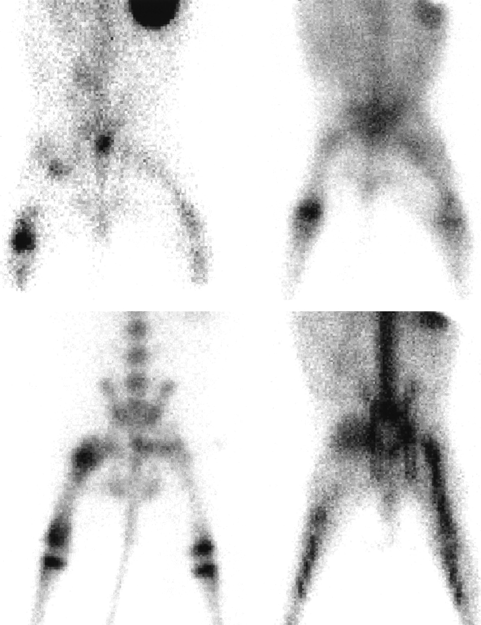
Scintigraphic analysis was performed on the images of all rabbits. Examples of the scintigraphic recordings are shown in Figure 3. All imaging agents correctly detected the acute osteomyelitis. The diaphyseal infection of the femur was best visualized with the delayed bone scan (Fig. 3, bottom, left) and, to a lesser extent, with 67Ga-citrate (Fig. 3, top, right), whereas the images with 99mTc-IL-8 and 111In-granulocytes were photopenic in the diaphysis. In contrast, infected areas at the proximal and distal parts of the femur were more clearly visualized with 99mTc-IL-8 and 99mTc-MDP. Equivocal results were found with 111In-granulocytes in 5 animals at 24 h after injection. False-positive findings were obtained with 99mTc-MDP (rabbit 3), based on delayed images only. A fracture of the epiphyseal–metaphyseal endplate of the proximal femur was found to be radiologically responsible for the increased osteoblastic uptake. In all infected rabbits investigated, 99mTc-IL-8 scintigraphy delineated the osteomyelitic lesion as early as 2 h after injection and the quality of image improved from 2 to 4 h after injection. Uptake of 67Ga-citrate and 111In-granulocytes in the osteomyelitic lesions allowed detection of the infectious focus only at 48 h after injection, as shown in Figure 4.

Scintigraphic images of rabbits with complete infection of femur. (Top) 99mTc-IL-8 image 4 h after injection (left) and 67Ga-citrate image 48 h after injection (right) for rabbit 8 of group B. (Bottom) 99mTc-MDP image 4 h after injection (left) and 111In-granulocyte image 48 h after injection (right) for rabbit 1 of group A.

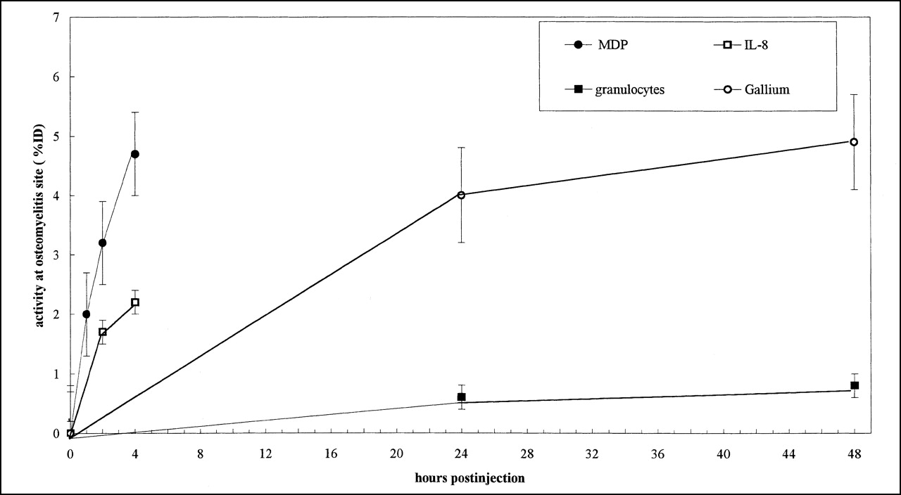
Uptake at osteomyelitic site as determined by quantitative analysis of scintigraphic images of group A rabbits administered 99mTc-MDP and 111In-granulocytes and group B rabbits administered 99mTc-IL-8 and 67Ga-citrate. Error bars represent SEM.
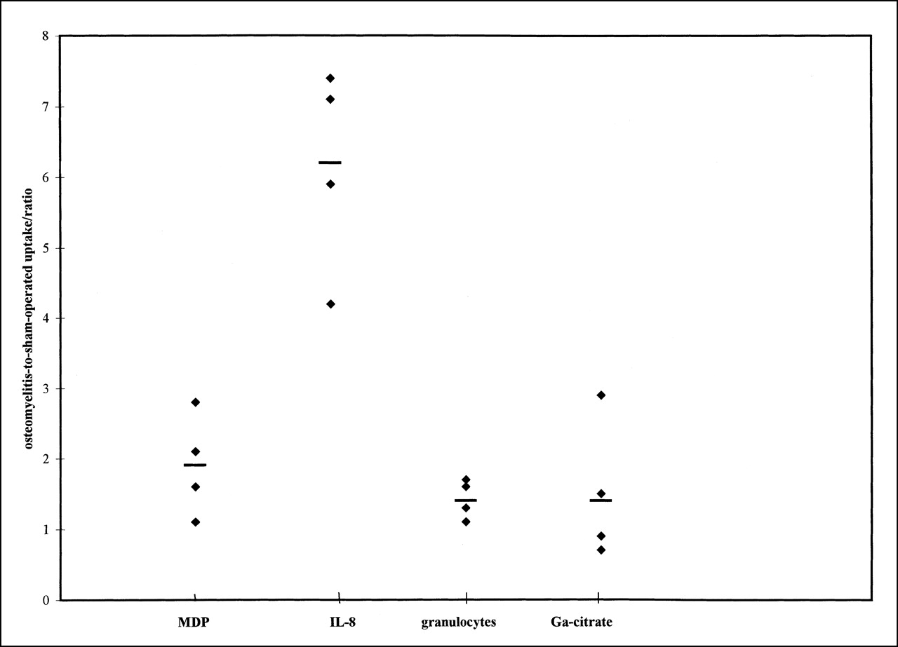
Uptake in the osteomyelitic lesion as a fraction of the whole-body activity was significantly higher for 99mTc-MDP and 67Ga-citrate than for the other 2 agents (P < 0.005). The bone-seeking agents 99mTc-MDP and 67Ga-citrate gave similar absolute uptake at all times (up to 4.7 ± 0.7 %ID and 4.9 ± 0.8 %ID, respectively). Lower absolute uptake values were found for 99mTc-IL-8 (up to 2.2 ± 0.2 %ID) and 111In-granulocytes (up to 0.8 ± 0.2 %ID). Uptake of 99mTc-IL-8 in the osteomyelitic lesion at 4 h after injection was, on average, 2.8-fold higher than that of 111In-granulocytes at 48 h after injection (Fig. 4). As shown in Figure 5, the T/Bs were similar for 99mTc-MDP (1.9 ± 0.2 at 4 h after injection), 67Ga-citrate (1.5 ± 0.4 at 48 h after injection), and 111In-granulocytes (1.4 ± 0.1 at 48 h after injection). The T/Bs obtained with 99mTc-IL-8 increased with time up to 4 h after injection (5.0 ± 0.1 at 2 h after injection and 6.2 ± 0.3 at 4 h after injection; P < 0.13, not significant). For 99mTc-IL-8 at 4 h after injection, the T/Bs were 3.5-, 4.4-, and 4.7-fold higher than the T/Bs of 99mTc-MDP at 4 h after injection, 67Ga-citrate at 48 h after injection, and 111In-granulocytes at 48 h after injection, respectively. The mean T/B of 99mTc-IL-8 (6.2 ± 0.3 at 4 h after injection) was significantly higher than the mean T/Bs of 67Ga-citrate, 99mTc-MDP, and 111In-granulocytes (1.5 ± 0.4, 1.9 ± 0.2, and 1.4 ± 0.1, respectively; P < 0.0001).

T/Bs calculated from quantitative region-of-interest analysis of scintigraphic images. Data are for 3-h (99mTc-MDP) and 48-h (111In-granulocyte) images for rabbits of group A and for 4-h (99mTc-IL-8) and 48-h (67Ga-citrate) images for rabbits of group B. Horizontal bars represent mean values.
DISCUSSION
The low diagnostic yield of radiography in this study confirmed that this technique is unreliable in establishing acute bone infection when an additional pathologic condition is present. In osteomyelitis, the bone destruction caused by vascular ischemia, by enzymes of disintegrated polymorphs, and by increased intramedullary pressure (32) can be visualized with bone-seeking agents such as 67Ga-citrate and 99mTc-MDP. Both radiopharmaceuticals have proven to be sensitive in detecting pathologically increased bone turnover, although the specificity of both agents is limited: other conditions may also cause increased uptake, such as tumors, activated osteoarthritis, and noninfectious inflammatory lesions (33). An increase in specificity can be achieved using agents targeting the neutrophils that have infiltrated the bone marrow. Up to now, these neutrophilic infiltrates have been visualized with radiolabeled leukocytes (34) or with radiolabeled antibodies directed against epitopes as present on granulocytes (35).
With 99mTc-IL-8, most infections in the rabbits were detected as early as 2 h after injection, whereas delayed 48-h postinjection images were necessary with 111In-granulocytes (36). With 99mTc-IL-8 as well as with 67Ga-citrate, all infected sites were detected, whereas delayed bone scanning alone was falsely positive in 1 rabbit and 111In-granulocytes missed 1 case of osteomyelitis.
This study showed that 99mTc-IL-8, a proinflammatory chemotactic cytokine, allows visualization of osteomyelitic lesions as evidenced by infiltrated neutrophils, presumably by targeting the surface receptors on granulocytes. Furthermore, 99mTc-IL-8 performed at least as well as 99mTc-MDP and 67Ga-citrate in the localization of acute bone infection. Although absolute uptake of the agent in the osteomyelitic lesion was lower, the T/B obtained with 99mTc-IL-8 was highest. The direct comparison with 111In-granulocytes showed superior targeting of 99mTc-IL-8.
The moderate performance of 111In-granulocytes in acute osteomyelitis is in line with studies on patients with osteomyelitis (37). The cause of the photopenic areas in infected bones imaged with 111In-granulocytes (in humans) is considered to be multifactorial and includes impaired blood supply and substitution of the bone marrow by pathologic processes. It has been documented that 14% of acute osteomyelitic lesions appear as cold lesions on scintigraphic images (38), most likely because subperiosteal and intraosseous pus compresses the microcirculation of the involved bone. Predominantly, this compression is thought to occur mainly in the femoral diaphysis and femoral head (39).
Granulocytes abundantly express IL-8 receptors (16–19) on their cell membrane. Moreover, IL-8 and tumor necrosis factor are locally produced by inflammatory cells in the bone marrow and play an important role in the attraction of neutrophils, which may result in a further increase in IL-8 receptor–positive cells in the infectious focus (1). The data in this study suggest that 99mTc-IL-8 is more suited than 111In-granulocytes for imaging osteomyelitis, presumably because infiltration of the radiolabeled granulocytes is hampered by the physiologic factors mentioned above (40).
The absolute uptake of MDP in the femur in the instance of false positivity (rabbit 3) was as high as 2.3 %ID and in the same range as the osteomyelitic cases, illustrating the increased uptake of 99mTc-MDP in any site of bone repair of any cause. Similarly, with 67Ga-citrate both the absolute uptake and the T/B were similar to those obtained with 99mTc-MDP. The values of the absolute uptakes in the infected bone were lower for 99mTc-IL-8 than for the bone-seeking agents, but the T/Bs were 4.7-fold higher for 99mTc-IL-8 than for the others. This finding indicates that there was no falsely positive uptake at the sham-surgery site and that 99mTc-IL-8 uptake increased at the infected site over time while the background activity decreased.
CONCLUSION
In this study, 99mTc-IL-8 accurately revealed acute osteomyelitis in a rabbit model. 99mTc-IL-8 correctly identified all rabbits with acute osteomyelitis within 4 h after injection. The ease of preparation, early good image quality, and lower radiation burden suggest that 99mTc-IL-8 may be a suitable imaging agent for the scintigraphic evaluation of acute osteomyelitis.
ACKNOWLEDGMENTS
The authors thank Gerry Grutters and Henny Eikholt (Central Animal Laboratory, University of Nijmegen) for expert assistance with the animals, Emile Koenders (Department of Nuclear Medicine, University Medical Center Nijmegen) for excellent technical assistance, and Drs. Pius Loetscher and Marco Baggiolini (Theodor Kocher Institute, University of Bern, Bern, Switzerland) for the kind gift of the cell lines. This study was supported in part by the Deutsche Forschungs Gesellschaft.
Footnotes
Received Jan. 16, 2001; revision accepted Apr. 9, 2001.
For correspondence or reprints contact: Stefan Gratz, MD, Department of Nuclear Medicine, University Medical Center Nijmegen, P.O. Box 9101, 6500 HB Nijmegen, The Netherlands.


