


Abstract
A novel, facile procedure for efficient coupling of high doses of 131I to monoclonal antibodies (MAbs) was developed with minimal chemical and radiation damage. Methods: To diminish the radiation and chemical burden during labeling, iodination was performed in a large reaction volume and by temporarily coating the MAb with a minimal amount of IODO-GEN. The MAb was coated by injection of IODO-GEN (dissolved in acetonitrile [MeCN]) into the aqueous MAb solution, and the coating was subsequently removed by addition of ascorbic acid. For chemoprotection before, during, and after PD-10 purification of the 131I-MAbs, ascorbic acid and human serum albumin were used. The effects of autoradiolysis in the starting 131I solution were countered by treatment with NaOH and ascorbic acid. For this so-called IODO-GEN–coated MAb method, the sensitive chimeric MAb MOv18 (c-MOv18) and the more robust murine MAbs K928 and E48 were used. The high-dose 131I-labeled MAbs were characterized for radiochemical purity and MAb integrity by thin-layer chromatography, high-performance liquid chromatography, and sodium dodecyl sulfate polyacrylamide gel electrophoresis followed by phosphor imager quantification. The high-dose 131I-labeled MAbs were also characterized for immunoreactivity. The radiopharmacokinetics and biodistribution of 131I-c-MOv18 were analyzed in human tumor–bearing nude mice. For comparison, 131I-c-MOv18 batches were made using the conventional chloramine-T or IODO-GEN–coated vial method. Results: Conventional high-dose labeling of 5 mg c-MOv18 with 4.4 GBq 131I resulted in a labeling yield of 60%, a radiochemical purity of 90%, an immunoreactive fraction of 25% (72% being the maximum in the assay used), and the presence of aggregation and degradation products. Using similar amounts of 131I and MAb in the IODO-GEN–coated MAb method, 85%–89% overall radiochemical yield, at least 99.7% radiochemical purity, and full preservation of MAb integrity and immunoreactivity were achieved. For this labeling, 5 mg MAb were coated with 35 μg IODO-GEN during 3 min in a reaction volume of 6 mL. Also, biodistribution was optimal, and tumor accumulation was superior to that of coinjected 125I-c-MOv18 labeled according to the conventional IODO-GEN–coated vial method. Conclusion: A new, facile, high-dose 131I-labeling method was developed for production of 131I-labeled MAbs with optimal quality for use in clinical radioimmunotherapy.
To come to a more effective treatment of ovarian cancer, we are focusing on the development of radioimmunotherapy (RIT). Radiolabeled monoclonal antibodies (MAbs) binding to tumor-associated antigens have been shown to have therapeutic efficacy in preclinical studies (1–3) as well as in several clinical studies (4–9).
On the road to tailoring targeted RIT, the availability of a MAb with appropriate specificity is a prerequisite (10). A candidate MAb for RIT of ovarian cancer is MAb MOv18. MOv18 binds to the membrane folate receptor, a 38-kDa glycoprotein, which is highly expressed on ovarian carcinoma cells, whereas expression on normal cells is much more restricted (11–13). The murine and chimeric forms (c-MAb) of MOv18 have been studied extensively in vitro, in animal models, and in ovarian cancer patients (4,5,14).
131I is still the most widely used radionuclide for RIT. It has an appropriate half-life and is a γ and β emitter that can be used for radioimmunoscintigraphy as well as for therapy. Importantly, 131I is readily available, and methods for coupling 131I to MAbs are easy. The maximum tolerated dose for 131I-labeled MAb IgG typically is approximately 3.7 GBq, but for myeloablative protocols this dose can be as high as 22.2 GBq (15).
High-dose 131I-MAb labeling for RIT is mostly performed with electrophilic iodine generated in situ by chloramine-T (15–17) or by 1,3,4,6-tetrachloro-3α,6α-diphenylglycouril (IODO-GEN; Pierce, Oud Beijerland, The Netherlands) using IODO-GEN–coated vials (18–21). A problem met in high-dose 131I labeling is impairment of the immunoreactivity of the MAb in association with altered biodistribution characteristics. This damage is the combined result of the high radioactivity concentration and the high amount of oxidant present during labeling. One can hypothesize that MAb damage will be reduced when labeling is performed in a larger volume (less radiation damage), using less oxidant (less chemical damage) and applying optimal chemoprotection. A challenge in the development of such a labeling procedure, however, is to retain high labeling yields.
In this article, we describe a novel, facile procedure for high-dose 131I labeling of MAbs. The procedure harbors three essential elements to preserve MAb immunoreactivity, integrity, and radiopharmacokinetics. First, before the start of the iodination, the 131I solution is pretreated to neutralize the oxidative effects of aging. Second, iodination is performed by temporarily coating the MAb with IODO-GEN. This iodination method reconciles the strongly conflicting demands of reducing the amount of IODO-GEN and increasing the reaction volume. Third, before, during, and after purification, protectants are used to further reduce potential MAb damage. High-dose 131I-c-MOv18 preparations obtained using this novel IODO-GEN–coated MAb procedure were characterized for MAb integrity and immunoreactivity and compared with 131I-c-MOv18 preparations made using the conventional IODO-GEN or chloramine-T method. Moreover, the biodistribution of low- and high-dose 131I-c-MOv18, prepared by the novel method, was evaluated in tumor-bearing nude mice with coinjected 125I-c-MOv18 labeled by the conventional IODO-GEN–coated vial method as a reference.
MATERIALS AND METHODS
MAbs
Production and characterization of chimeric MOv18 have been described elsewhere (4,22). Purified c-MOv18 IgG for human use was provided by Centocor B.V. (Leiden, The Netherlands). The murine control MAbs m-K928 and m-E48, binding to squamous cell carcinomas, have been described elsewhere (23,24).
High-Dose Labeling with Conventional IODO-GEN–Coated Vial Method
A representative iodination protocol was used (20). Briefly, a 2-mL solution containing 4.4 GBq 131I (7.4 GBq/mL, 10 μg 127I/mL; Amersham, Aylesbury, U.K.) and 5 mg c-MOv18 was added to a glass vial coated with 500 μg IODO-GEN. After wobbling for 10 min, the reaction mixture was passed through a 0.22 μmol/L MillexGV filter (Millipore, Etten-Leur, The Netherlands) and purified on a PD-10 column (Pharmacia, Roosendaal, The Netherlands) with 0.9% NaCl as the equilibration solution and the eluent. Aliquots of the product were taken for thin-layer chromatography (TLC), high-performance liquid chromatography (HPLC), sodium dodecyl sulfate polyacrylamide gel electrophoresis ([SDS-PAGE] reducing and nonreducing conditions), and phosphor imager quantification and for determination of the immunoreactive fraction.
High-Dose Labeling with Conventional Chloramine-T Method
Two representative iodination protocols were used. Method 1 included a 2-mL solution containing 4.4 GBq 131I, 10 mg c-MOv18, and 200 μg chloramine-T 3H2O (N-chloro-p-toluenesulfonamide, sodium salt, trihydrate; Merck, Darmstadt, Germany), which was incubated for 3 min (17). After being quenched with 200 μg Na2S2O5, the iodinated MAb was purified and analyzed as described above. In method 2, the reaction was performed in 2 mL with 4.4 GBq 131I, 5 mg c-MOv18, and 600 μg chloramine-T 3H2O, with a reaction time of 3 min (20); quenching was performed with 800 μg Na2SO3.
For evaluation of the potential damaging effect of Na2SO3, 0.5 mL of a crude iodination reaction mixture (1 mg MAb in 1 mL; 185 kBq 131I; reaction time, 5 min; 75 μg vial-coated IODO-GEN) was incubated with either 1 mg Na2SO3 or 1 mg ascorbic acid (control) during 5 min. After PD-10 purification, the products were analyzed as described above.
High-Dose Labeling with Novel IODO-GEN–Coated MAb Method
Pretreatment of 131I Activity.
Before the delivery vial was opened, the gaseous activity was removed by a 50-mL syringe (for quantification purposes) or a sterile suction pump. The content of the delivery vial was adjusted to 1 mL with 1 mmol/L NaOH and transferred to a 20-mL β-scintillation glass vial, and 10 μL ascorbic acid (80 nmol, pH 5) were added. Samples taken from vials (3.7 GBq/500 μL) before and 1 min after the addition of NaOH and ascorbic acid were analyzed by HPLC. For labeling of MAbs, 200 μL 1 mol/L Na2HPO4 (pH 7.2) were added 1 min after the ascorbic acid addition to bring the pH of the 1.2-mL starting 131I solution to 7.2.
Temporary Coating of MAb with IODO-GEN.
The coating of MAb molecules with IODO-GEN and subsequent removal of the coating with ascorbic acid were visualized by ultraviolet (UV) detection (315 and 280 nm) during HPLC analysis. Samples (20 μL) were analyzed from native MAb (2.5 mg MAb in 1.0 mL 0.2 mol/L phosphate buffer, pH 6.8), coated MAb (after addition of 25 μL IODO-GEN/acetonitrile [MeCN], 10 mg/mL), and MAb with coating removed (after subsequent addition of 25 μL ascorbic acid, 100 mg/mL). The removal of the coating by ascorbic acid (reduction of IODO-GEN to 3α,6α-diphenylglucouril) has been monitored in situ in a nuclear magnetic resonance (NMR) tube in a 9:1 (volume per volume) mixture of CD3CN and D2O. NMR data: Ascorbic acid: d, 1H, δ 4.73 (J = 2.0 Hz); m, 1H, δ 3.84 (J = 2.0, 6.3, and 6.9 Hz); m, 2H, δ 3.56 (J = 11.3, 6.9, and 6.3 Hz). Dehydroascorbic acid: d, 1H, δ 4.52 (J = 0.8 Hz); m, 1H, δ 4.37 (J = 0.8, 3.3, and 5.6 Hz); dd, 1H, δ 4.16 (J = 10.0 and 5.6 Hz); dd, 1H, δ 4.00 (J = 10.0 and 3.3 Hz). IODO-GEN: m, 8 H, δ 7.20 (ortho + meta H); dd, 2H, δ 7.00 (para H; J = 8.2 and 1.5 Hz). 3α,6α-Diphenylglycouril: m, 10H, δ 7.18–7.07.
Assessment of Labeling and Purification Conditions.
The new coating approach was evaluated by several sets of labeling experiments within the following general frame: every reaction was performed with a pretreated 131I solution that contained 80 nmol ascorbic acid, irrespective the amount of 131I; the added MAbs (1 mg) were dissolved in 0.1 mol/L phosphate buffer, pH 6.8, with the final pH of the reaction mixture being 7.0–7.2; the reaction was started by injection of the chosen aliquot of IODO-GEN/MeCN (stock: 1 mg/mL) into the solution and abrogated with 100 μL ascorbic acid (25 mg/mL, pH 5).
In the first set, the labeling efficiency was assessed in a reaction volume of 2, 6, and 12 mL for the MAbs c-MOv18, m-K928, and m-E48 using 37 MBq 131I, 75 μg MAb-coated IODO-GEN, and a reaction time of 5 min. For a comparison, this set of labeling experiments was also performed with 75-μg IODO-GEN–coated vials. Labeling percentages were assessed with TLC, whereas MAb integrity was evaluated by HPLC.
In the second set, the amount of IODO-GEN coated to the MAb was varied (50, 35, 25, and 15 μg), using a reaction volume of 2 mL, 37 MBq 131I, the MAbs c-MOv18 and m-K928, and a reaction time of 5 min. Labeling percentages were assessed with TLC.
In the third set for all three MAbs, the labeling kinetics were evaluated for 35 μg MAb-coated IODO-GEN in a reaction volume of 2, 6, and 12 mL, using 37 MBq 131I. Aliquots of 10 μL were taken at 1, 2, 3, 4, and 5 min. Each aliquot was put into a vial containing 25 μL ascorbic acid solution (25 mg/mL) to stop the labeling. Labeling percentages were assessed by TLC.
In the fourth set, postiodination processing conditions were evaluated, taking the radiochemical purity and the MAb integrity as a measure. Coating was performed with 35 μg IODO-GEN, using 1.7 GBq 131I, a reaction time of 5 min, a reaction volume of 2 mL, and the MAbs c-MOv18, m-K928, and m-E48. The 131I-MAbs were purified on PD-10 columns with 0.9% NaCl containing 5 mg/mL ascorbic acid (pH 5.0) as the equilibration solution and eluent. Before the PD-10 purification, either no or 50 μL 20% human serum albumin (HSA) were added to the reaction mixture; after the purification either no or 20% HSA was added (1% [weight per weight] final concentration). The 131I-MAbs were analyzed by TLC and SDS-PAGE (reducing and nonreducing conditions) followed by phosphor imager quantification. Stored samples (1-mL solutions containing 0.37 GBq 131I) at room temperature were analyzed by TLC during 50 h.
High-Dose Labeling.
The MAbs c-MOv18, m-K928, and m-E48 were iodinated in 6 mL, using 5 mg MAb, 2.2 GBq 131I, and a reaction time of 3 min, essentially according to the protocol in Table 1. For determination of the radiophysical limit, the 131I dose was increased with stepwise increments of 0.7 GBq in the case of c-MOv18. Postiodination processing was performed as described in steps 8–11 (Table 1). The labeling procedure was performed under good manufacturing practice (GMP) conditions with the aid of an automated labeling device, and all reagents were sterile and pyrogen free. The 131I-MAbs were analyzed by TLC, HPLC, and SDS-PAGE, and the immunoreactive fraction was determined. For a final analysis of the overall in vitro stability, 5 mg 131I-c-MOv18 (740 MBq/mg MAb) in 10 mL 0.9% NaCl, 5 mg/mL ascorbic acid, and 10 mg/mL HSA, pH 5.0, were stored in a 25-mL glass vial at room temperature. TLC and HPLC were performed after 0.1, 1, 2, 4, and 24 h. Gel electrophoresis and measurement of the immunoreactivity were performed at 1 and 24 h.
Technical Protocol for High-Dose 131I Labeling According to IODO-GEN–Coated MAb Method
Iodinated MAb Preparations for Biodistribution Experiments
Low- and high-dose 131I-c-MOv18 was labeled according to the new method in 6 mL, starting with 352 MBq (final specific activity, 66.6 MBq/mg) and 4.3 GBq (final specific activity, 762 MBq/mg). 125I-c-MOv18 was labeled according to the IODO-GEN–coated vial protocol used in previous patient studies (14). Briefly, 55.5 MBq 125I (1.48 GBq/mL; Amersham) in 600 μL sterile phosphate-buffered saline (PBS), pH 7.4, were added to 1 mL c-MOv18 (1 mg), mixed, and incubated for 6 min at room temperature in a vial coated with 100 μg IODO-GEN. Free iodine was removed by a sterile anion-exchange resin in PBS for 1 min (Dowex AG1-X8; BioRad, Utrecht, The Netherlands) in the presence of 10 μg KI, and the reaction mixture was passed through a 0.22-μm MillexGV filter. A radiochemical purity similar to that of the new method was obtained through additional purification by PD-10 column (eluent: 0.9% NaCl, 5 mg/mL ascorbic acid, pH 5.0), resulting in a final radiochemical purity of 99.5% (TLC) and a specific activity of 37 MBq/mg.
Analyses
For HPLC analysis of the 131I activity in the delivery vial, an RP-Select B column (125 × 4 mm, 5 μm; Merck) was used. The eluent was 0.05 mol/L NaH2PO4·H2O/0.002 mol/L (Bu)4NH4OH (pH 7), and the flow rate was 0.8 mL/min. The HPLC retention times were 1.7 min for IO3− and 5.0 min for I−. Identification of the other 131I species was not attempted.
The proton NMR (1H NMR) spectra of ascorbic acid, dehydroascorbic acid, IODO-GEN, and 3α,6α-diphenylglycouril were recorded in CD3COOD or CD3CN:D2O (9:1 [volume per volume]) on an AC 200 (200.13 MHz) spectrometer (Bruker, Coventry, U.K.). Chemical shifts are given in δ (ppm) relative to δ (CD2HCN) = 1.93.
For determination of the PD-10 column elution profile of 3α,6α-diphenylglycouril by HPLC, an RP18 Chromspher C18 column (250 × 4.6 mm, 5 μm; Chrompack, Middelburg, The Netherlands) was used. The eluent was H2O:EtOH 60:40 (volume per volume), and the flow rate was 0.8 mL. The HPLC retention time of 3α,6α-diphenylglycouril was 6.4 min. The PD-10 column elution profile of MeCN has been determined previously (25).
Analyses of the radioiodinated MAbs by HPLC, TLC, and gel electrophoresis followed by phosphor imager quantification were performed as described recently (26). HPLC retention times were 22 min for 131I-MAb IgG and 40 min for free 131I species. Complexes of high molecular weight eluted at 18–20 min. The immunoreactivity of radioiodinated MAbs was determined in a cell-binding assay on the respective target cells as described previously (4). For c-MOv18, the human nasopharyngeal epidermoid carcinoma cell line KB, expressing high levels of membrane folate receptor, was purchased from Flow Laboratories (Herts, U.K.). KB cells were grown as a monolayer in folate-free Roswell Park Memorial Institute tissue culture medium and 10% dialyzed fetal calf serum (Life Technologies, Paisley, U.K.) using subphysiologic concentrations of folate to upregulate the receptor. The cells were washed with PBS and bovine serum albumin (1%) just before use. For the control MAbs m-E48 and m-K928, UM-SCC-22B cells were used (27). The optimal immunoreactive fractions of c-MOv18, m-E48, and m-K928 were 72%, 95%, and 82%, respectively.
Biodistribution
Nude mice bearing subcutaneous human ovarian cancer xenografts, IGROV1, in the left and right abdominal side were used as described earlier (4). Thyroid uptake was blocked by the addition of potassium iodide to the drinking water (0.1%) starting 3 d before the experiment. Tumor-bearing animals (mean tumor weight ± SD, 0.24 ± 0.19 g) received intravenous injections of a mixture of 125I-c-MOv18, 131I-c-MOv18, and unlabeled c-MOv18, and the syringe was weighed before and after injection. The mice were killed at 3, 6, and 24 h after injection; four mice were used for each time point. Blood was collected under ether anesthesia. Normal tissues and tumors were then dissected, rinsed in saline to minimize blood residues, dried, and weighed. The radioactivity was measured in a γ well counter with automatic correction for the 131I Compton effect in the 125I window setting. For calculation of the injected dose, six weighed standard solutions of the injected material were prepared and counted simultaneously with the tissues. The results were expressed as percentage injected dose per gram.
Differences in tissue uptake between coinjected MAbs were statistically analyzed for each time point with SPSS 7.5 software (SPSS Inc., Chicago, IL) using the Student t test for paired data. Two-sided significance levels were calculated, and P < 0.05 was considered statistically significant.
RESULTS
High-Dose Labeling with Conventional Methods
High-dose labeling of c-MOv18 with the IODO-GEN–coated vial method resulted in an overall yield of 60%, a radiochemical purity of 91%, and 25% immunoreactivity. Phosphor imager analysis of the SDS-PAGE gel (Fig. 1) revealed, besides the presence of the major 150-kDa IgG band (peak 3) and free iodine (peak 7), the presence of aggregation products (peaks 1 and 2) and products of smaller molecular weight (shoulder at peak 3 + peaks 4, 5, and 6). Analysis of a 186Re-c-MOv18 preparation as a control revealed only the 150-kDa peak. Under the same labeling conditions, a low-dose labeling of c-MOv18 with 18.5 MBq 131I gave 6% aggregation products, whereas immunoreactivity was found to be 48%, indicating a significant chemical contribution of the oxidant to the overall damage. Furthermore, when the same radioactivity concentration (2.2 GBq/mL) was applied during labeling of m-K928 and m-E48, the integrity was affected accordingly. The drop in immunoreactivity was, in this case, approximately 35% for both MAbs.

Phosphor imager profile of SDS-PAGE gel of 131I-c-MOv18, high-dose–labeled with conventional IODO-GEN–coated vial method. Quantification report: 1, 1.1%; 2, 8.6%; 3, 70.4%; 4, 8.7%; 5, 1.2%; 6, 1.1%; 7, 8.9%. Conditions: 4.4 GBq 131I, 5 mg c-MOv18, 500 μg IODO-GEN, 2-mL reaction volume, 10-min reaction time.
On high-dose labeling of c-MOv18 with chloramine-T as the oxidant, the results did not improve. The overall labeling yield was also ±60%, the immunoreactive fraction measured 29% (method 1) and 19% (method 2), and impairment of the integrity was similar to that shown in Figure 1. In addition, an increased peak at approximately 100 kDa was observed, most probably caused by the quenching procedure with the sulfite-based reducing agents (20). For verification, we incubated a 131I-c-MOv18 preparation with Na2SO3 before PD-10 purification. Phosphor imager analysis revealed that the reducing agent indeed affected the integrity of the MAb (18% of the radioactivity co-migrating with a 100-kDa peak). However, immunoreactivity was also reduced (to 58%), indicating that such sulfite-based reducing additives are not innocuous in two aspects. Analogous incubation with ascorbic acid did not affect the integrity and gave an immunoreactive fraction of 72%.
Labeling with Novel IODO-GEN–Coated MAb Method
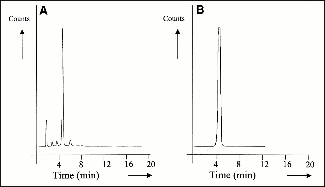
From 7 to 11 MBq 131mXe and from 18 to 37 MBq gaseous 131I were measured in a 131I delivery vial containing 3.7 GBq. Therefore, sucking off the gaseous phase before opening the delivery vial is recommended. Monitoring of the air inside the laminar flow hood showed that formation of gaseous activity during the subsequent experiments was negligible. Analysis of the 131I solution in the delivery vial revealed that, because of the radiolysis, the pH had dropped from 11 to 7–9 and that a proportion of the 131I had been oxidized to 131IO3− (retention time, 1.7 min) and three additional products (Fig. 2A) that cannot form carbon–iodine bonds. Adjustment of the pH of the 131I solution, transfer of the radioactivity from the blackened delivery vial to the reaction vial, and addition of 80 nmol ascorbic acid as the reducing agent resulted in a solution free from radiolysis products, as was judged from the regeneration of the 131I into a mono 131I− peak (Fig. 2B). Introduction of this procedure made all 131I recovered from the delivery vial (95%–96%) available for the labeling reaction.

HPLC profile of 131I quality of aged 131I solution in blackened delivery vial (3.7 GBq in 500 μL) 4 d after production (A) and after adjustment of pH, transfer to reaction vial, and addition of ascorbic acid (B). Percentages: (A) 13% 131IO3− (retention time [Rt], 1.7 min), 79% 131I− (Rt, 5.0 min), 8% unidentified 131I components. (B) 100% 131I− (Rt, 5.0 min).
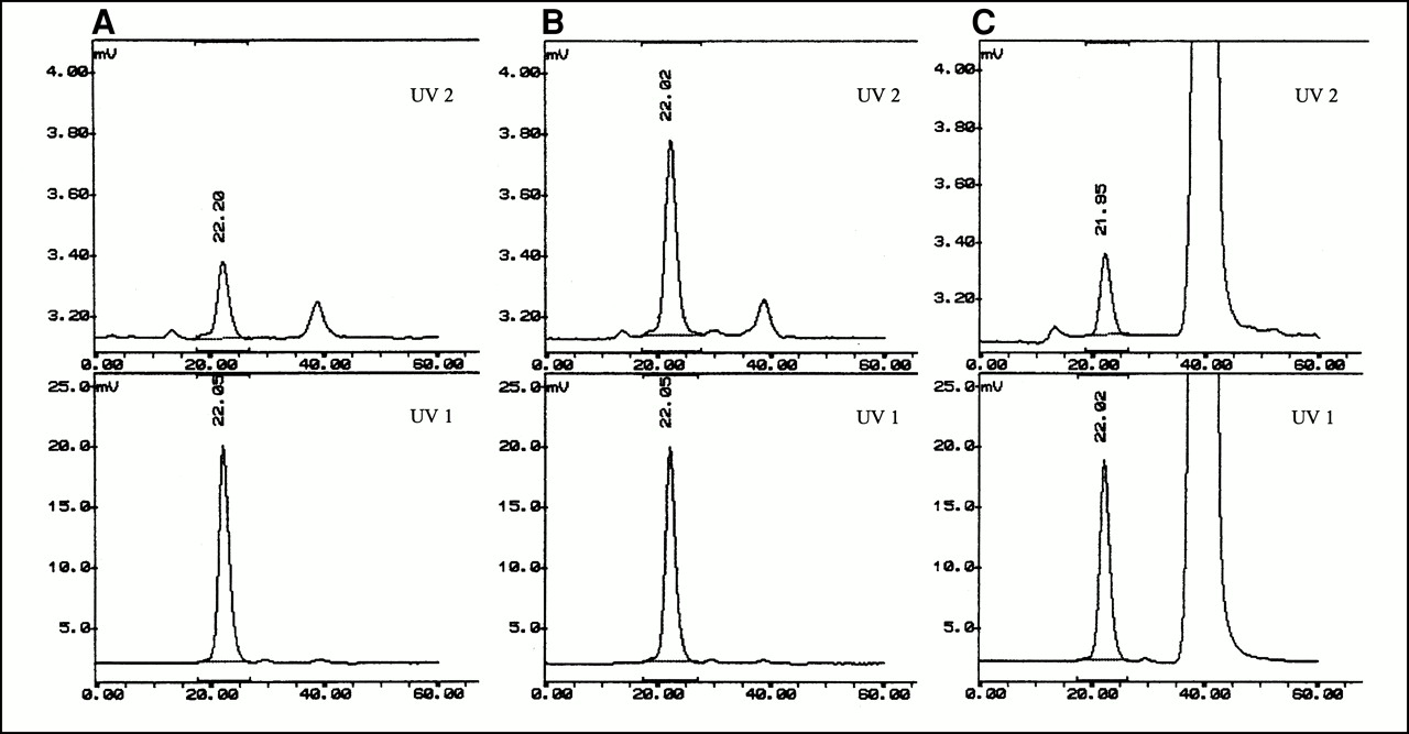
To dislodge the kinetic implacability of labeling MAbs with 131I under conditions of large reaction volume and small amounts of oxidant, the MAb was temporarily coated with IODO-GEN. The coating process is shown in Figure 3 using UV-detectable amounts of IODO-GEN. On addition of IODO-GEN dissolved in MeCN, the IODO-GEN “precipitates” on the apolar parts of the MAb molecules (Fig. 3B, top panel). This coating appeared to be a random process, as was shown using a mixture of MAb and bovine serum albumin. On addition of ascorbic acid, IODO-GEN is detached from the macromolecules by reduction of the IODO-GEN into the corresponding 3α,6α-diphenylglycouril compound (Fig. 3C). Addition of 131I to this solution did not result in labeling of the MAb, indicating that all IODO-GEN had become inactivated. Assessment of the PD-10 elution profile of 3α,6α-diphenylglycouril by HPLC (detection at 210 nm) and 1H NMR analysis revealed that the compound was quantitatively collected in fractions eluted after the MAb-containing fractions.

HPLC visualization of temporary coating of antibody with IODO-GEN and subsequent removal of coating by ascorbic acid through simultaneous UV detection at 315 nm, second absorption maximum of IODO-GEN (UV 2), and, at 280 nm, absorption maximum of MAb (UV 1). Shown are native MAb (A), coated MAb (B), and MAb with coating removed (C). Conditions are described in text. Large peak at 40 min (C) is from ascorbic acid; minor peaks (at 16 and 39 min) in chromatograms (A and B) at 315 nm are solvent peaks.
Using 37 MBq pretreated 131I and 1 mg MAb coated with 75 μg IODO-GEN, the new approach showed, after 5 min, greater than 90% labeling yield for the three MAbs, even in a volume of 12 mL. In contrast, using the IODO-GEN–coated vial method, after 5 min the yields were 25% ± 3%, 9% ± 2%, and 4% ± 2% in a reaction volume of 2, 6, and 12 mL, respectively, illustrating the strongly improved contact between the IODO-GEN and the reactants in the new procedure. HPLC analysis gave one monomeric MAb peak at 22 min, indicating that coating 1 mg MAb during 5 min with this amount of IODO-GEN did not lead to adverse oxidative aggregation. When the amount of IODO-GEN was decreased (in search of a further reduction of chemical burden), 35 μg IODO-GEN was found to give a labeling yield of greater than 90% after 5 min. Kinetic measurements with this amount of IODO-GEN in 2, 6, and 12 mL showed, independent of the MAb used, greater than 90% labeling after 2, 3, and 5 min, respectively. Within this stoichiometry (Table 1, footnote), a large flexibility regarding the specific activity of 131I-MAb could also be achieved, at least from a chemical point of view: starting with 1.7 GBq 131I, a product with a specific activity of 1.5 GBq/mg MAb was obtained. However, the MAb integrity of such a product was not preserved without protection.
The most effective protection against radiation damage after iodination, as well as on subsequent storage, was accomplished by the introduction of HSA and ascorbic acid (Table 1, steps 8–11). Under these protection conditions, the challenging 1.7-GBq reactions resulted in a radiochemical purity greater than 99%, whereas phosphor imager quantification revealed the best preservation of MAb integrity (150-kDa band, 92% ± 3%). Subsequent storage of labeled MAb for 50 h as a 0.37 GBq 131I/mL sample resulted in a deiodination of 0.08%/h. Without the two HSA steps, the radiochemical purity was 96%–97% and the deiodination was 0.27%/h; without the HSA and ascorbic acid in the PD-10 eluent, the radiochemical purity was 90%–93% and the deiodination was 1.10%/h. SDS-PAGE under reducing conditions followed by phosphor imager quantification showed that the heavy chain–versus–light chain 131I ratio was 8:1 in the case of MOv18 and 2:1 in the case of K928 and E48. With the conventional IODO-GEN–coated vial method, the same ratios were found.
The final consideration was the overall quality of the MAb, including preservation of immunoreactivity. High-dose 131I-MAb preparations were made under the established conditions (Table 1) using 2.2–4.3 GBq 131I. With this procedure, overall radiochemical yields were 85%–89%, whereas the radiochemical purity immediately after purification was greater than 99.7%. One hour after labeling with 2.2 GBq, the immunoreactivity and integrity of the three MAbs were optimal. The procedure allowed an increase to 4.3 GBq 131I, as shown by SDS-PAGE and phosphor imager analysis for MAb c-MOv18 (Fig. 4A). The immunoreactivity measured 72%, which is also optimal for this MAb. Because 740 MBq 131I contain 1 μg 127I, the resulting 740 MBq/mg MAb corresponds to an overall iodine-to-MAb ratio of 1.36 and an 131I-to-MAb ratio of 0.18, which means that roughly one of five MAb molecules carries an 131I atom.

Phosphor imager profile of SDS-PAGE gel of 131I-c-MOv18, 1 h (A) and 24 h (B) after formulation. Radioactivity concentration: 0.37 GBq/mL. Quantification report: (A) 1, 0.4%; 2, 98.3%; 3, 1.3%. (B) 1, 1.5%; 2, 80.1%; 3, 10.9%; 4, 2.7%; 5, 1.4%; 6, 3.3%. Conditions (IODO-GEN–coated MAb method): 4.3 GBq 131I, 5 mg c-MOv18, 35 μg IODO-GEN, 6-mL reaction volume, 3-min reaction time.
Samples were stored for 24 h at room temperature and analyzed for overall in vitro stability. Under the most challenging storage conditions (0.37 GBq 131I/mL, 0.5 mg MAb/mL), ascorbic acid and HSA could not fully protect the MAb against the direct hits of the β-particles. After 24 h, the integrity of the MAbs was affected in the way shown in Figure 4B for c-MOv18; in this case, the radiochemical purity decreased to 96.7% and the immunoreactivity dropped to 62%. These quality aspects improved when conjugates were diluted (with 0.9% NaCl, ascorbic acid, and HSA) to 0.18 and 0.09 GBq 131I/mL before storage.
Biodistribution Studies
MAb c-MOv18, labeled with a high and low dose of 131I according to the novel IODO-GEN–coated MAb method, was analyzed in nude mice bearing subcutaneous human ovarian cancer xenografts, with coinjection of a low-dose 125I-c-MOv18 labeled according to the conventional IODO-GEN–coated vial method. Mice received either 5 μg high-dose 131I-c-MOv18 (3.8 MBq) coinjected with 5 μg 125I-c-MOv18 (185 kBq) or 5 μg low-dose 131I-c-MOv18 (333 kBq) coinjected with 5 μg 125I-c-MOv18 (185 kBq). By addition of unlabeled c-MOv18, the total injected amount of c-MOv18 was 50 μg in each case.
The immunoreactivity measured 72% for the high- and low-dose 131I-c-MOv18 and 68% for the 125I-c-MOv18 (Fig. 5). Judging from the steepening of the slope, the avidity of the 125I-c-MOv18 preparation seemed slightly less than that of the two 131I-c-MOv18 preparations.

Immunoreactivity assay of radioiodinated c-MOv18 MAbs used for biodistribution analysis: 125I-c-MOv18 (▪); 131I-c-MOv18, high dose (▴); 131I-c-MOv18, low dose (•). T/C values (T = total radioactivity; C = activity specifically bound to cells) are function of inverse of cell concentration.
Biodistribution analysis was performed at 3, 6, and 24 h after injection. The results obtained for the high-dose series are compiled in Table 2. The 131I liver accumulation data at 3 h revealed a significantly (P = 0.020) increased hepatic extraction (4.3% ± 0.3% [131I] vs. 3.8% ± 0.3% [125I]). For all other normal organs, the 131I and 125I data were fully congruent throughout the period studied, whereby the low 131I stomach accumulation data at 3 h (0.7% ± 0.2% for both isotopes) were in accordance with the absence of free contaminating iodide in the product. Interestingly, all 24 tumors (8 tumors per group, 2 tumors per individual mouse) showed a significant 131I/125I ratio above 1. At 3 and 6 h, this ratio was 1.05–1.10 (P = 0.031 and P = 0.007, respectively); at 24 h, this ratio was increased to 1.15–1.20 (P = 0.001). This 15%–20% higher 131I tumor accumulation at 24 h was accompanied by an increased disappearance of 131I label from the blood (P = 0.071); this approximately 10% decrease tended to be related to the size of the tumor.
Biodistribution Data of IGROV1 Tumor-Bearing Nude Mice After Coinjection of High-Dose Labeled 131I-c-MOv18 and Low-Dose Conventionally Labeled 125I-c-MOv18
The data of the parallel low-dose experiment were found to agree fully with the data shown in Table 2, with one exception: the liver accumulation data of the 3-h group were the same (3.0% ± 0.3% [131I] vs. 2.9% ± 0.3% [125I]).
DISCUSSION
During the past few decades, the possibility of using radiolabeled MAbs for radioimmunoscintigraphy and RIT has been intensively investigated. For many RIT studies, 131I was considered the radionuclide of choice because it has an appropriate physical half-life (8 d), β-particle energy (Emax = 0.6 MeV), and pathlength (r90 = 0.83 mm, r90 being the range in which 90% of the energy is released) and its γ emission permits imaging and absorbed dose calculations. Because more 131I-labeled MAbs are entering phase I, II, and III clinical RIT trials, the need for high-dose labeling methods that are easy and safe to perform under GMP conditions has increased. High-dose labeling demands minimization of chemical and radiation damage to MAbs, because such damage results in impairment of the immunoreactivity and integrity of the MAb, accompanied by altered pharmacokinetics and suboptimal tumor targeting.
In this article, we describe a novel method for high-dose 131I labeling of MAbs. Essential to this procedure is temporary coating of the MAb with IODO-GEN, enabling the use of larger reaction volumes. The labeling procedure appeared highly efficient (overall labeling yield > 85%) and resulted in conjugates with high radiochemical purity (>99%), optimal integrity, and optimal immunoreactivity for specific activities up to 740 MBq 131I/mg MAb. Also, optimal biodistribution characteristics were obtained as assessed in tumor-bearing nude mice for c-MOv18, a MAb that became heavily damaged when conventional high-dose 131I labeling methods (chloramine-T and IODO-GEN–coated vial methods) were applied.
In a high-dose 131I-labeling procedure, the first critical step is the iodination itself. During the labeling reaction, no antioxidants are allowed to minimize the radiation-induced deterioration of the MAb. The option to spread both the chemical and the radiation damage over a larger number of MAb molecules is not realistic, because the resulting higher MAb concentration will suffer more direct hits, and the time delay between formation and reaction of radiolysis products with the MAb will be shortened. The only legitimate option, therefore, is to apply the dilution principle with respect to the MAb concentration as well as the radioactivity concentration, using a large reaction volume combined with a short reaction time and a small amount of oxidant.
The harsh oxidant chloramine-T was found to be unsuitable when used in small amounts in a large reaction volume. Although ascorbic acid appeared to be an adequate alternative for Na2SO3 or Na2S2O5 to quench the reaction, the intrinsic nonselectivity of chloramine-T resulted in suboptimal quality of the 131I-c-MOv18 and too low radiochemical yields. After the indirect chloramine-T labeling route using an active ester (28,29), ascorbic acid could not be used as a quenching agent because it destroyed the active ester. Furthermore, in this more laborious procedure, solubility problems were met, and a moderate overall radiochemical yield is inherent to the method.
The conventional IODO-GEN–coated vial method is also notoriously inefficient in larger reaction volumes because of the inappropriate contact area between the IODO-GEN on the glass wall and the reactants in the solution (131I and the MAb). Two attempts to enhance contact between IODO-GEN and the reactants using an aqueous IODO-GEN suspension have been reported (30,31). However, these approaches did not lead to a practical and applicable high-dose 131I-labeling methodology. Therefore, to speed up the reaction, one was compelled to increase the amounts of vial-coated IODO-GEN to 500–1000 μg (20,21,32,33). Nevertheless, the radiochemical yields were only moderate (20,21,32) and sometimes unreliable (21). In addition, as shown in this study, a serious chemical contribution to the overall damage can be introduced by such high IODO-GEN–to–MAb molar ratios, a problem left unnoticed when picomolar amounts of MAb were used (34).
In the IODO-GEN–coated MAb method, IODO-GEN and MAb are temporarily in closest proximity to each other in solution, so that contact with the third component, the 131I atoms in solution, is the only reaction rate parameter. This approach offered the desired labeling kinetics within the framework of a large reaction volume and a small amount of oxidant.
Two additional radiation damage–related adjustments were made, namely pretreatment of the starting 131I solution and the use of ascorbic acid and HSA after iodination. Similar measures were also successfully implemented in a high-dose 186Re labeling protocol (25,27). In that protocol, Na2SO3 could be used to counter the effects of aging of the 186Re solution, because this reagent did not face the MAb (removal by Sep-Pak [Waters Corp., Milford, MA] before conjugation). However, in a one-pot labeling, the use of an agent that affects both the integrity and the immunoreactivity of the MAb should be avoided. Ascorbic acid was found to be a perfect alternative for Na2SO3. After addition to the starting 131I solution, remaining ascorbic acid protects the added MAb until addition of the IODO-GEN. The implication is that some of the added IODO-GEN will be reduced. However, this minor inactivation was considered when assessing the amount of IODO-GEN to be used in the labeling procedure. In the optimized postiodination processing, ascorbic acid removes the coating from (reduction of IODO-GEN) and regenerates (reduction of potentially formed sulfonium chloride bonds) the MAb and provides chemoprotection (reduction of the formed radiolysis products). HSA acts as a buffer against directs hits of the MAb molecule by the β-particles and assists in restoring possible disturbances of the folding of the MAb after and during the ascorbic acid regeneration process. The presence of ascorbic acid in the eluent provides the necessary chemoprotection during PD-10 purification of the 131I-MAb/HSA mixture. Accordingly, the radiochemical purity of the final product is also strongly improved by suppressing the deiodination, which is one of the manifestations of radiation damage.
Regarding protection after purification, cryopreservation to retard the reaction of radiolysis products with the MAb has been reported as an option (33). However, without an encapsulating agent, freezing of a solution does not affect the chance of direct hits of the MAb molecule by β-particles, and without an antioxidant, any remaining and newly formed reactive species will attack the MAb during and after thawing. Other investigators (15,17,20,35) applied the principle of dilution (to 37–74 MBq/mL) in combination with HSA as antioxidant and encapsulating agent. We prefer the combination of ascorbic acid and HSA because this antioxidant mixture is more potent and therefore allows more concentrated radioimmunoconjugate solutions.
For 5 mg MAb coated with 35 μg IODO-GEN in a reaction volume of 6 mL (i.e., 0.8 mg MAb/mL), the radiophysical limit of the IODO-GEN–coated MAb method was found to be 0.74 GBq 131I/mL, resulting in 3.7 GBq 131I-MAb with an overall iodine-to-MAb ratio of 1.36. The preservation of immunoreactivity at this ratio leads to the important conclusion that a relationship between impairment of immunoreactivity and the iodine-to-MAb molar ratio, if any, does not exist up to this radiophysical limit. Moreover, the fact that with the conventional IODO-GEN–coated vial method a nearly indifferent 131I-c-MOv18 batch was obtained using the same amounts of radioactivity and MAb clearly shows that the radiation and the high IODO-GEN–to–MAb molar ratio, not the introduction of an iodine atom into the antigen binding site of the MAb, were responsible for the observed drop in immunoreactivity (16,34,36–39). This finding strongly suggests that the underlying nature of decreased immunoreactivity and avidity, as caused by the radiation and the oxidant, is impaired folding or impaired flexibility of the antigen binding sites. This impairment may be caused by changed locoregional polarity caused by affected S–S bridges and oxidized amino acids such as methionine and tryptophan. This concept is well in line with our demonstrated impairment of immunoreactivity by oxidant and Na2SO3. For both iodinating agents, a specific way to chemically affect sulfur atoms is the formation of intermediary sulfonium chloride bonds. Without a regeneration step, these bonds hydrolyze to polar SOH or S–O bonds or lead to aggregation, and quenching with SO32− may lead to formation of polar bonds such as S–SO3−.
Biodistribution analysis revealed that the radiopharmacokinetics of 131I-c-MOv18 that was labeled with a specific activity of 762 MBq/mg MAb using the new method fully paralleled that of accordingly labeled 131I-c-MOv18 with a specific activity of 67 MBq/mg MAb and also that of 125I-c-MOv18, made by the conventional IODO-GEN–coated vial method. Hence, labeling a MAb to an iodine-to-MAb ratio of 1.36, corresponding to one to three iodine atoms per IgG, does not implicate a radiopharmacokinetic restriction.
Conventionally labeled 125I-c-MOv18 exhibited impaired immunoreactivity and avidity in comparison with 131I-c-MOv18 obtained by the new method. This finding implies that, in IODO-GEN–coated vial labeling, the chemical burden onto 1 mg MAb exceeded the combined radiation and chemical burden onto 5 mg MAb in the new procedure. The subtle difference in immunoreactivity and avidity of the 125I-c-MOv18 was reflected in 15%–20% less activity in the tumor at 24 h after injection. Given the great impact of this seemingly close difference in quality and the fact that a greater than 20% drop in immunoreactivity is not uncommon for conventionally high-dose–labeled 131I-MAbs, it seems that in those studies in vivo deiodination or the release of 131I-tyrosine (in cases of internalizing MAbs (40)) have not been the major factors responsible for low tumor uptake and retention.
CONCLUSION
In 131I-RIT studies, optimal radiochemical purity, integrity, immunoreactivity, and avidity should have first priority, especially if various MAbs, dosing schedules, and radionuclides are compared. The IODO-GEN–coated MAb method provides a route to achieve this goal.
ACKNOWLEDGMENTS
The authors thank Dr. Wim den Hollander for labeling c-MOv18 with 125I. The continuous support of Dr. Sven O. Warnaar is much appreciated. This study was supported by the Dutch Cancer Society and the Dutch Medical and Health Research Organization NWO (Nederlandse Organisatie voor Wetenschappelijk Onderzoek).
Footnotes
Received Jul. 5, 2000; revision accepted Nov. 10, 2000.
For correspondence or reprints contact: Gerard W. Visser, PhD, Radionuclide Center Vrije Universiteit, De Boelelaan 1085C, 1081 HV Amsterdam, The Netherlands.
REFERENCES
In this issue


Jump to section
Related Articles
Cited By...
- Development of Novel ADCs: Conjugation of Tubulysin Analogues to Trastuzumab Monitored by Dual Radiolabeling
- Phase 0 Microdosing PET Study Using the Human Mini Antibody F16SIP in Head and Neck Cancer Patients
- IDENTIFICATION OF INFLAMED ATHEROSCLEROTIC PLAQUE USING 123I-LABELED INTERLEUKIN-2 SCINTIGRAPHY IN HIGH-RISK PERITONEAL DIALYSIS PATIENTS: A PILOT STUDY
- In-Line Radiolabeling: A Novel Continuous-Flow System for Commercial-Scale Protein Labeling
- Improved tumor targeting of anti-epidermal growth factor receptor Nanobodies through albumin binding: taking advantage of modular Nanobody technology
- Radioimmunotherapy of Head and Neck Cancer Xenografts Using 131I-Labeled Antibody L19-SIP for Selective Targeting of Tumor Vasculature
- 89Zr as a PET Surrogate Radioisotope for Scouting Biodistribution of the Therapeutic Radiometals 90Y and 177Lu in Tumor-Bearing Nude Mice After Coupling to the Internalizing Antibody Cetuximab
- 131I-Rituximab: Relationship Between Immunoreactivity and Specific Activity
- 89Zr Immuno-PET: Comprehensive Procedures for the Production of 89Zr-Labeled Monoclonal Antibodies