


Abstract
Serotonin-1A (5-hydroxytryptamine-1A [5-HT1A]) receptors have been reported to play an important role in the pathophysiology of a variety of psychiatric and neurodegenerative disorders. Animal experiments have shown that 4-(2′-methoxyphenyl)-1-[2′-(N-2′′-pyridinyl)-p-[18F]fluorobenzamido]ethylpiperazine ([18F]MPPF) may be suitable for 5-HT1A receptor imaging in humans. The aim of this study was to determine if [18F]MPPF can be used for the quantitative analysis of 5-HT1A receptor densities in brain regions of healthy human volunteers. Methods: [15O]H2O perfusion scanning was performed before intravenous injection of [18F]MPPF to obtain anatomic information. Cerebral radioactivity was monitored using a PET camera. Plasma metabolites of [18F]MPPF were determined by high-performance liquid chromatography. Binding potentials were calculated using the metabolite-corrected arterial input function and a linear graphic method (Logan-Patlak analysis). Results: The highest levels of radioactivity were observed in the medial temporal cortex, especially in the hippocampal area. In contrast, the cerebellum and basal ganglia showed low uptake of 18F, in accordance with known 5-HT1A receptor distribution. The calculated binding potentials correlated well with literature values for 5-HT1A receptor densities. The binding potentials for [18F]MPPF were 4–6 times lower than those that have been reported for [carbonyl-11C]-(N-(2-(4-(2-methoxyphenyl)-1-piperazinyl)ethyl)-N-(2-pyridyl) cyclohexanecarboxamide (WAY 100635), indicating that [18F]MPPF has a lower in vivo affinity for 5-HT1A receptors. Conclusion: These results confirm that [18F]MPPF can be used for the quantitative analysis of 5-HT1A receptor distribution in the living human brain. The rapid dissociation from the receptor makes this ligand a possible candidate to monitor changes in endogenous serotonin levels.
- human
- brain
- serotonin-1A receptors
- 5-hydroxytryptamine-1A receptors
- 4-(2′-methoxyphenyl)-1-[2′-(N-2′′-pyridinyl)-p-fluorobenzamido]ethylpiperazine
During the last decade, it has become clear that serotonin-1A (5-hydroxytryptamine-1A [5-HT1A]) receptors play an important role in the pathophysiology of a variety of psychiatric and neurodegenerative disorders (1–5). Although in vitro autoradiography studies have reported changes in receptor density for schizophrenia, depression, and dementia (6–12), how these changes are related to in vivo functionality is still unclear. PET has the unique ability to quantitatively monitor physiologic changes in living tissue. Recently, several radioligands have been developed for the imaging and quantification of 5-HT1A receptors with PET, and several of these have been tested in humans (13–16). Especially, [carbonyl-11C]-(N-(2-(4-(2-methoxyphenyl)-1-piperazinyl)ethyl)-N-(2-pyridyl) cyclohexanecarboxamide ([carbonyl-11C]WAY 100635) has been reported to bind with high affinity (inhibition constant [Ki] = 0.8 nmol/L) (17) to the 5-HT1A receptor and to give good target-to-nontarget ratios (18–20). However, the high affinity of this tracer may restrict its use for measuring changes in endogenous serotonin levels. The selective 5-HT1A antagonist 4-(2′-methoxyphenyl)-1-[2′-(N-2′′-pyridinyl)-p-fluorobenzamido]ethylpiperazine (MPPF) has a somewhat lower affinity for the 5-HT1A receptor (Ki = 3.3 nmol/L) (17) and may therefore be more suitable for the detection of changes in endogenous serotonin. Recently, MPPF has successfully been labeled with [18F]fluorine, resulting in the [18F]fluoro analog [18F]MPPF (21). Animal experiments have shown a regional distribution of this radioligand that agrees well with known 5-HT1A receptor densities (21,22) and correlates well with autoradiography data (23).
The objective of this preliminary PET study was to determine if [18F]MPPF can be used for the quantitative analysis of 5-HT1A receptor densities in brain regions of healthy human volunteers. A recently developed method to determine regional distribution volumes through linear graphic analysis (23) was used to calculate the binding potentials, using the cerebellum as reference tissue because this region is practically devoid of 5-HT1A receptors (Eq. 1) (24,25).
 This method has already been applied to the analysis of [11C]raclopride studies (26) and has also been used for quantitative analysis of the in vivo binding of [carbonyl-11C]WAY 100635 to 5-HT1A receptors in the human brain (19,20). Because the binding potential corresponds to receptor density (Bmax)/dissociation constant (KD) (Eq. 1), it is a measure of both receptor density and the affinity of the radioligand for its receptor.
MATERIALS AND METHODS
Volunteers
The study was approved by the medical ethics committee of Groningen University Hospital. Five volunteers (2 men, 3 women; age range, 21–65 y) were included after written informed consent had been obtained and an independent physician had confirmed that they were healthy. Suitability to take part in the study was determined by compliance with the following criteria: an age between 18 and 65 y; a healthy status according to medical examination; no history of neurodegenerative or psychiatric disorders; no use of drugs such as neuroleptics, sedatives, or antidepressants; no use of corticosteroids or agents that suppress adrenal function; and, for women, no pregnancy or possibility of pregnancy.
Radiochemistry
[18F]MPPF was prepared by nucleophilic substitution of the aromatic nitro group as had been done previously (J. Passchier, unpublished data, 1998; (21,22) describe a comparable method). Quality control was performed using reverse-phase high-performance liquid chromatography (Nova-Pak, 150 × 3.9 mm; Waters, Milford, MA). A 28:6:65 mixture of acetonitrile:tetrahydrofuran:0.01 N NaOAc was used, pH was 5, retention time (tR) ([18F]MPPF) was 5 min, and tR (nitro precursor of MPPF) was 7 min. The levels of the nitro precursor were << 1 mg/L, and no detectable amounts of WAY-100634 (nonradioactive compound formed by hydrolysis of the nitroprecursor, with affinity for the 5-HT1A and α1-adrenergic receptors) (14,27) were observed. The radiochemical purity of the product was greater than 99%.
PET Experimental Procedure
PET was performed on an ECAT 962/HR+ camera (Siemens Medical Systems, Inc., Hoffman Estates, IL), giving 63 slices with a center-to-center distance of 2.425 mm. Full width at half maximum was 4–5 mm. Before the PET scan, the volunteers were positioned in the camera using a head restraint, and a transmission scan was obtained using 3 68Ge–68Ga rod-sources. After the transmission scan, the volunteers received an intravenous bolus injection of 1850 MBq [15O]H2O in 10 mL saline (0.9%). Radioactivity in the brain was measured during 15 min. Eight consecutive frames were acquired. Fifteen to thirty minutes after administration of [15O]H2O, during which time the volunteers remained in the camera, 79 MBq [18F]MPPF (specific activity > 110 TBq/mmol at the time of injection) in 10 mL phosphate buffer (Na3PO4 = 9.0 mmol/L; Na2HPO4 = 1.3 mmol/L; 0.9% saline) at pH = 7.4–7.6 with 7%–8% ethanol were administered intravenously as a bolus. Twenty-one consecutive frames were acquired: 6 frames at 10 s each, 2 frames at 30 s each, 3 frames at 60 s each, 2 frames at 120 s each, 2 frames at 180 s each, 3 frames at 300 s each, and 3 frames at 600 s each. Arterial blood samples were taken manually at the midpoint of each frame. The hematocrit level was determined. After plasma was acquired by centrifugation, samples of 250 μL were counted in a calibrated γ counter (Compugamma 1282 CS; LKB-Wallac, Turku, Finland) to obtain the arterial input function. Whole-blood samples (250 μL) were also counted to estimate uptake of [18F]MPPF in erythrocytes.
Metabolite Analysis
Arterial plasma samples (1 mL) were deproteinized with 70% perchloric acid (0.05–0.1 mL volume). After precipitation of protein by centrifugation, the supernatant was injected onto a reverse-phase high-performance liquid chromatography system (Novapak, 150 × 3.6 mm) A 45:55:0.3 mixture of MeOH:0.1 N NH4HCO2:Et3N was used, and the flow rate was 1.0 mL/min. Thirty samples were collected during 15 min using a fraction collector. The retention time of the parent compound was 9 min.
Plasma protein binding or free fraction of 18F was determined by the ultrafiltration technique, using a reusable micropartition system (MPS-1) with a molecular mass cutoff of 30,000 Da (regenerated cellulose membrane; Amicon, Beverly, MA). Samples of human plasma (0.25 mL) were dispensed into MPS-1 units and centrifuged at 2000g for 30 min. Radioactivity in the colorless ultrafiltrate and activity remaining on the filter were then determined by γ counting. A sample blank consisting of 300–500 Bq [18F]MPPF in saline was run in parallel; nonspecific adsorption to regenerated cellulose membranes was 7.5%.
Regions of Interest
The image obtained by summation of the [15O]H2O frames was used to obtain anatomic information for drawing regions of interest (ROIs), in particular for the cerebellum, striatum, and thalamus, in which uptake of [18F]MPPF was low. ROIs were drawn in the transaxial orientation using a contour tool (CAPP; Siemens/CTI, Knoxville, TN) for the cerebellum; striatum; thalamus; and frontal, cingulate, insular, lateral temporal, and medial temporal cortices. ROIs for the insular and medial temporal cortices were drawn directly on the summed [18F]MPPF image and not on the [15O]H2O image. Areas were located using a stereotactic atlas (28).
Calculation of Binding Potentials
Using the metabolite-corrected arterial input function and the ROI-derived time–activity curves, distribution volumes were calculated through linear graphic analysis (23). Binding potentials were estimated using Equation 1 (26). Because the blood volume in the brain was neglected (and is generally small, 3%–5%), binding potential = Bmax/KD (23).
RESULTS
Kinetics
[18F]MPPF showed rapid uptake in the brain followed by fast washout from the cerebellum and slower washout from target areas. Time–activity curves for the cerebellum and the frontal, lateral temporal, and medial temporal cortices are shown in Figure 1.

Time–activity curves for several brain regions after intravenous injection of 79 MBq [18F]MPPF in single subject.
Distribution
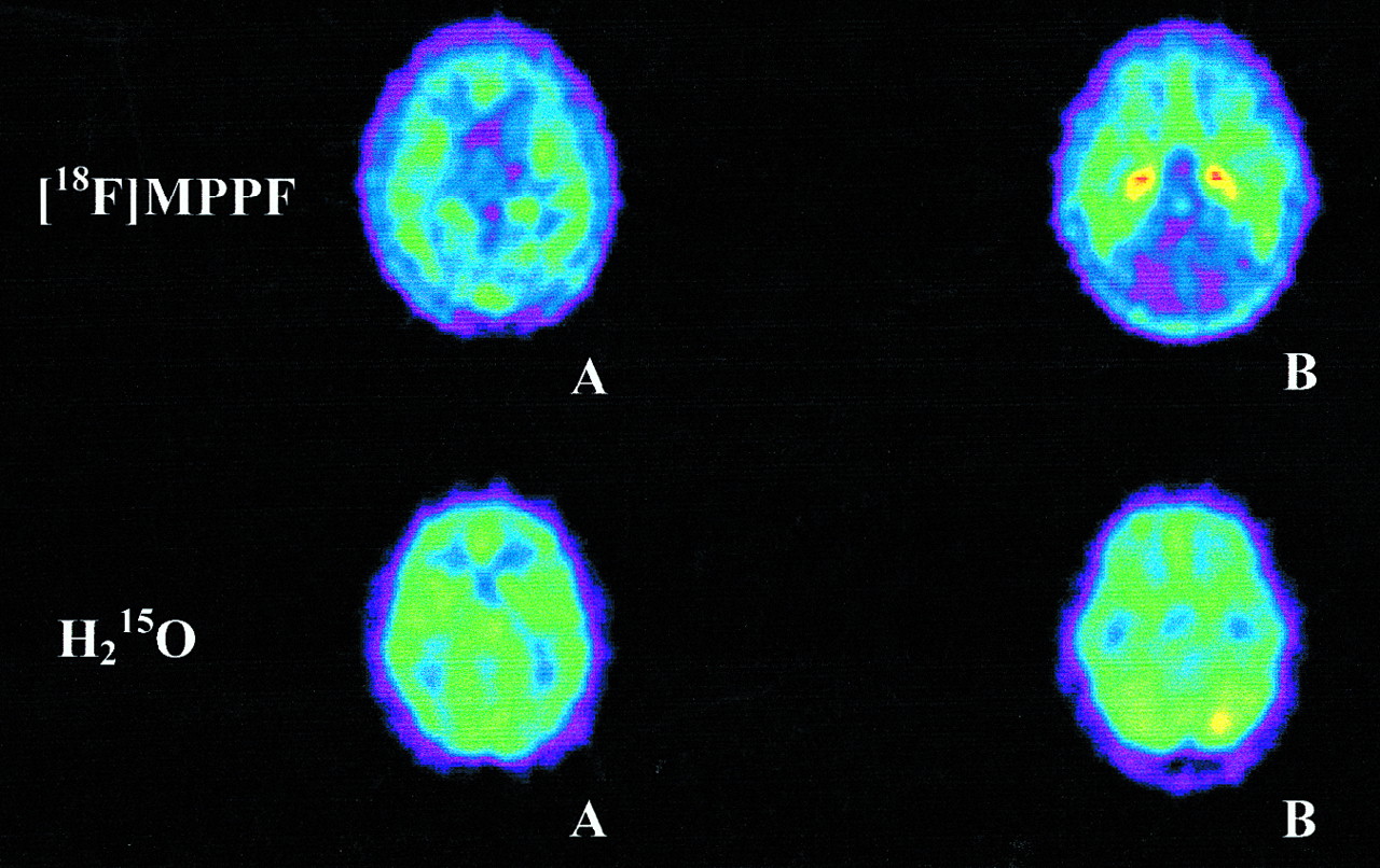
A comparison of the distribution of [18F]MPPF and [15O]H2O showed a clear difference in regional uptake (Fig. 2). Uptake of [18F]MPPF was low in the basal ganglia (Fig. 2A) and cerebellum (Fig. 2B), whereas high uptake was observed in the medial temporal cortex, especially in the hippocampal area (Fig. 2B). The flow tracer [15O]H2O, in contrast, showed high uptake throughout the brain, including the basal ganglia and cerebellum (Figs. 2A and B). The distinctive features of the hippocampal areas could not be observed in the [15O]H2O perfusion scan (Fig. 2B). Region-to-cerebellum ratios reached a maximum at 30–40 min after injection, with values ranging from 1.23 for the thalamus to 5.34 for the medial temporal cortex.

Comparison between distribution of [18F]MPPF and flow tracer [15O]H2O. For clarity, images were processed using gaussian filter (CAPP; Siemens/CTI). A = level of basal ganglia; B = level of medial temporal cortex.
Metabolism
Injected [18F]MPPF was rapidly metabolized (Fig. 3). After 10 min, only 10% of the radioactivity in the plasma represented parent compound. Analysis of [18F]MPPF-derived radioactivity in the plasma using reverse-phase high-performance liquid chromatography showed only 1 radioactive metabolite, with a retention time of 3 min (Fig. 4). A large fraction of injected [18F]MPPF (∼89%) was bound to plasma proteins. The radiolabeled metabolites of MPPF showed less protein binding (∼65%), a finding that is not surprising because they are more hydrophilic than the parent compound. When the free fraction of [18F]MPPF was arbitrarily manipulated in a Logan-Patlak analysis, binding to plasma proteins had no influence on the estimation of binding potential.
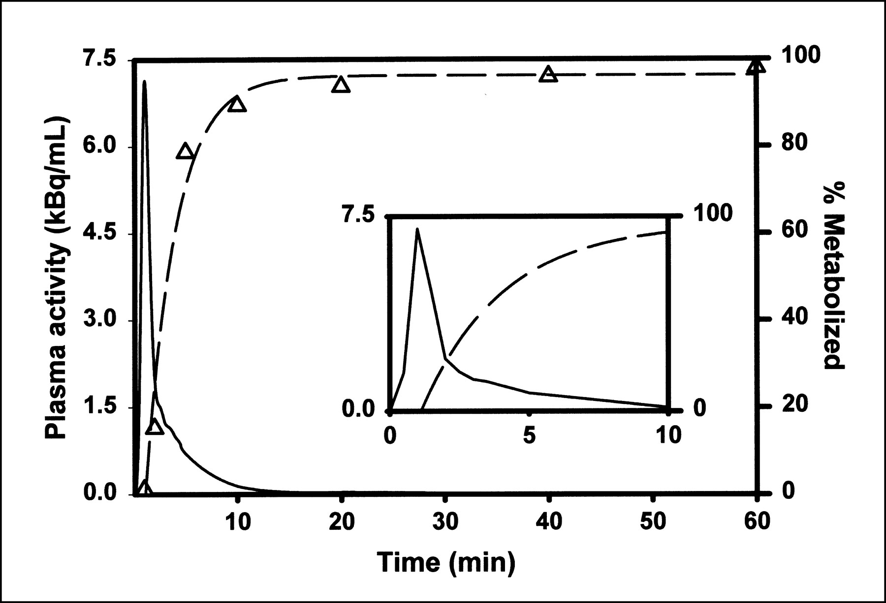
Rate of metabolism of [18F]MPPF (△ and dashed line represent fit) in single volunteer. Solid line represents metabolite-corrected arterial input function. Time interval from 0 to 10 min is shown in inset.
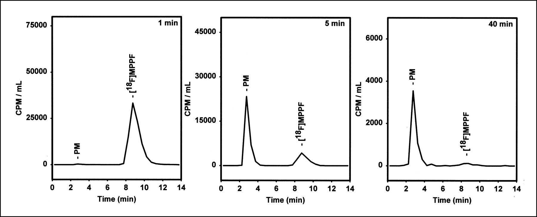
Radiochromatograms of [18F]MPPF-derived radioactivity in human plasma at 1, 5, and 40 min after injection. PM = polar metabolite.
Distribution Volumes and Binding Potentials
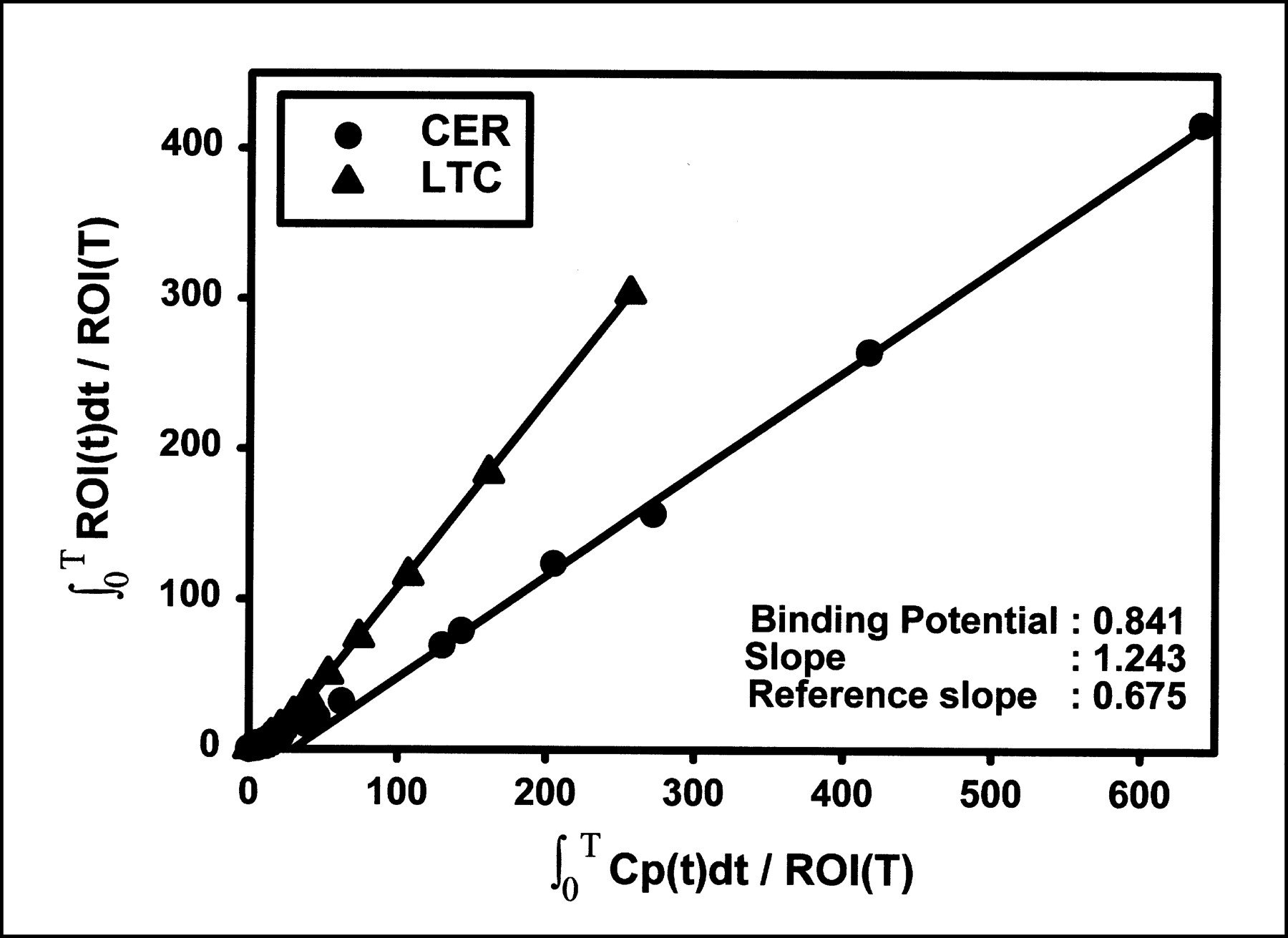
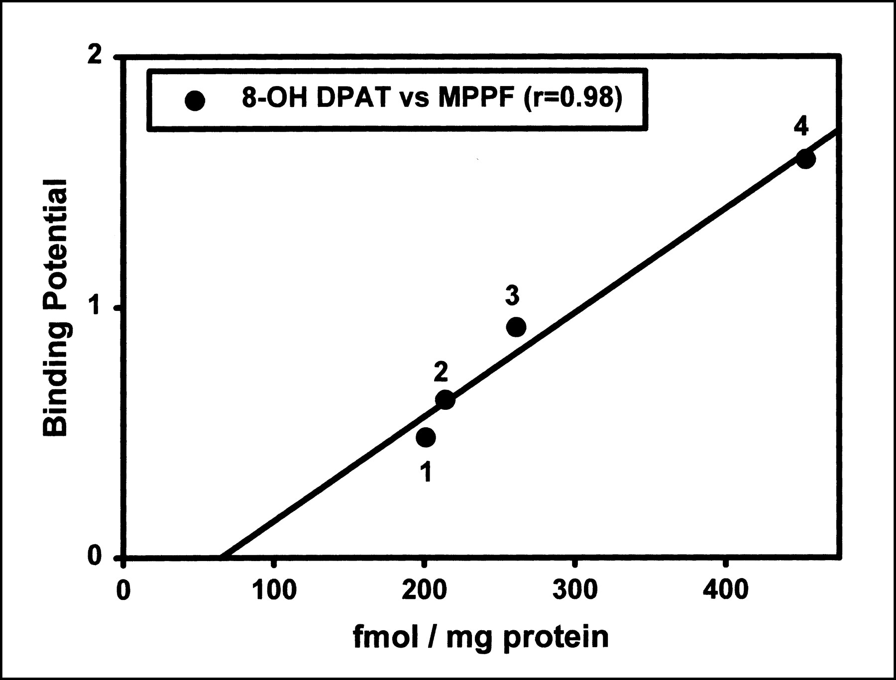
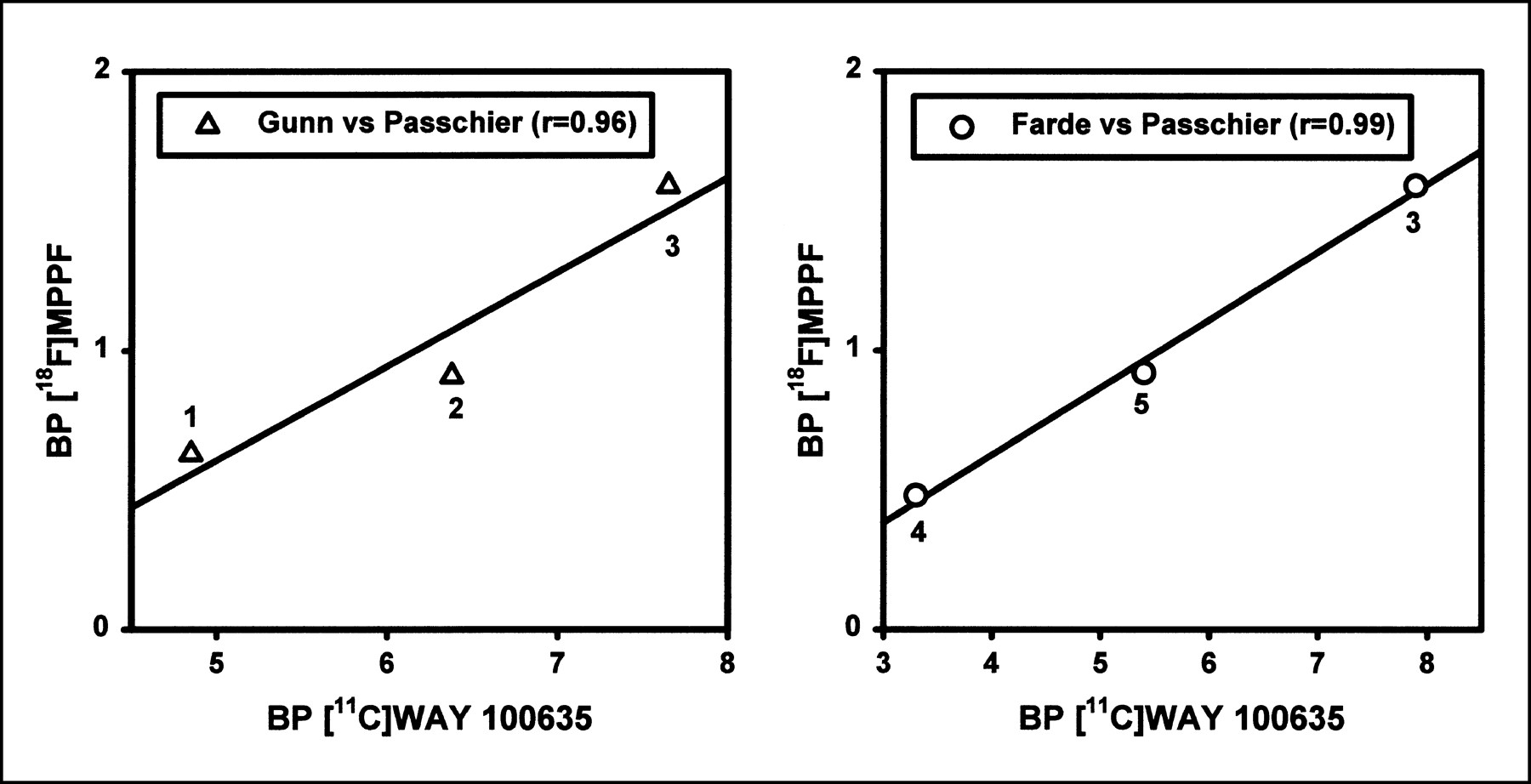
The metabolite-corrected arterial input (Fig. 3) was used to calculate distribution volumes from the ROI-derived regional time–activity curves by means of linear graphic analysis (Fig. 5) (23). Table 1 shows the results for several regions and their corresponding binding potentials. The calculated binding potentials (Eq. 1) correlated well with ex vivo quantitative autoradiography data for [3H]8-OH-2-(di-n-propylamino)tetralin (Fig. 6) and with reported values for [carbonyl-11C]WAY 100635 (Figs. 7A and B) (12,18,19).

Example of linear graphic analysis using Logan-Patlak fits. Slopes of fits represent the distribution volumes. ROI(T) refers to radioactivity in region of interest at time t. CER = cerebellum; Cp(t) = plasma radioactivity corrected for metabolites (23); ∫0T Cp(t)dt = integral of plasma radioactivity over time from time zero to time t; LTC = lateral temporal cortex; ∫0T ROI(t)dt = integral of radioactivity in region of interest over time from time zero to time t.

Correlation of distribution of [18F]MPPF in vivo with that of [3H]8-OH-2-(di-n-propylamino)tetralin in postmortem human brain (12). 1 = frontal cortex; 2 = cingulate cortex; 3 = insular cortex; 4 = medial temporal cortex (including hippocampal area).

Binding Potentials for Several Brain Areas
DISCUSSION
After intravenous administration of [18F]MPPF, a regional distribution was observed that differed markedly from that of the flow tracer [15O]H2O (Fig. 2) and corresponded to known 5-HT1A receptor localization (24,25) and with previous PET studies that used [carbonyl-11C]WAY 100635 (14,18–20). These results suggest that the distribution of 18F in the human brain after administration of [18F]MPPF reflects local 5-HT1A receptor density rather than blood flow.
Time–activity curves show rapid uptake of [18F]MPPF followed by fast washout from the cerebellum and slower washout from cortical areas (Fig. 1). The dissociation of [18F]MPPF from the receptor is clearly much more rapid than the dissociation of [carbonyl-11C]WAY 100635 (18–20), possibly because of the lower affinity of MPPF. This lower affinity results in lower image contrast, which may limit the use of MPPF for imaging applications. Especially small areas, such as the raphe nuclei, are hard to identify. When quantification of these regions is required, use of a higher affinity radioligand such as [carbonyl-11C]WAY 100635 may be necessary. Quantitative analysis of the binding of [18F]MPPF using a linear graphic method (24) with a metabolite-corrected arterial input function correlated well with ex vivo autoradiography using [3H]8-OH-2-(di-n-propylamino)tetralin (12) (Fig. 6) and with in vivo data for [carbonyl-11C]WAY 100635 (18–20) (Figs. 7A and B).
The rapid dissociation of [18F]MPPF, compared with the dissociation of [carbonyl-11C]WAY 100635, may enable measurement of changes in the endogenous serotonin concentration in the human brain brought about by, for example, selective serotonin reuptake inhibitors (29). The low inhibition constant of [18F]MPPF may also be useful for studying occupancy of 5-HT1A receptors by unlabeled drugs. Work is in progress to examine the possibility of estimating binding potential without the need for arterial sampling and metabolite analysis.
CONCLUSION
This study shows that [18F]MPPF can be used to delineate 5-HT1A receptors in healthy human volunteers using a linear graphical method. Even with low amounts of [18F]MPPF (79 MBq), a good correlation was found between regional [18F]MPPF uptake and 5-HT1A receptor densities. The rate of metabolism of [18F]MPPF was comparable with that of [carbonyl-11C]WAY 100635. The more rapid dissociation of MPPF may allow measurement of changes in endogenous serotonin concentration brought about in the brain by selective serotonin reuptake inhibitors and assessment of the occupancy of 5-HT1A receptors by (newly developed) unlabeled drugs.
Footnotes
Received Oct. 12, 1999; revision accepted Mar. 28, 2000.
For correspondence or reprints contact: Jan Passchier, PhD, PET Center, Groningen University Hospital, 9700 RB Groningen, The Netherlands.
REFERENCES
In this issue


Jump to section
Related Articles
Cited By...
- Species Differences in Blood-Brain Barrier Transport of Three Positron Emission Tomography Radioligands with Emphasis on P-Glycoprotein Transport
- Acute Treatment with the Antidepressant Fluoxetine Internalizes 5-HT1A Autoreceptors and Reduces the In Vivo Binding of the PET Radioligand [18F]MPPF in the Nucleus Raphe Dorsalis of Rat
- A Reduced Extracellular Serotonin Level Increases the 5-HT1A PET Ligand 18F-MPPF Binding in the Rat Hippocampus
- On the Quantification of [18F]MPPF Binding to 5-HT1A Receptors in the Human Brain