


Abstract
N-isopropyl-p-[123I]iodoamphetamine (IMP) SPECT is of low diagnostic value in patients with brain tumors, because brain tumors are visualized as uptake defects. Some reports have described non-Hodgkin's lymphoma of the central nervous system (CNS) as showing high uptake on delayed 123I-IMP SPECT images, suggesting its usefulness in diagnosing CNS lymphoma. In this study, we investigated the clinical value of 123I-IMP SPECT as a diagnostic tool for CNS lymphoma. Methods: Ninety-six patients with brain tumors, including 12 patients with CNS lymphoma, underwent 123I-IMP SPECT. Eleven patients had primary CNS lymphoma, and 1 had a parenchymal brain metastasis from a breast lymphoma. The total number of lesions was 18, 14 of which were in the cerebral parenchyma, 3 in the brain stem, and 1 in the ventricle. Early SPECT images were initiated 15–30 min after intravenous injection of 111 MBq 123I-IMP, and delayed images were collected 4 h later. SPECT images were visually analyzed with a color-grading scale. Tumor-to-normal activity ratio (T/N) and tumor-to-cerebellum activity ratio (T/C) were calculated for both early and delayed images for semiquantitative analysis. Results: By visual estimation, more than a 3- cm3 volume of CNS lymphoma was detected as an obvious focus of increased accumulation on delayed images. All other brain tumors tested appeared as decreased accumulation on delayed images. T/Ns and T/Cs on delayed images of CNS lymphomas, including tumors less than 3 cm3 in volume, were 1.48 ± 0.42 and 1.08 ± 0.16, respectively. These ratios in patients with glioma (0.30 ± 0.05 and 0.31 ± 0 07 respectively) or meningioma (0.34 ± 0.10 and 0.41 ± 0.17, respectively) showed a significant difference from those in patients with CNS lymphoma (P < 0.0005). Conclusion: 123I-IMP SPECT is a helpful tool for diagnosing CNS lymphoma.
Since the introduction of CT and MRI, the reported incidence of non-Hodgkin's lymphoma of the central nervous system (CNS) has increased (1). The incidence of primary CNS lymphoma, in particular, has increased, trebling in the last decade, partly because of an increase in the number of immunocompromised patients, including those with AIDS, those undergoing chemotherapy, and those with transplanted organs (1,2). Because of the characteristic appearance and localization of CNS lymphoma on CT scans and MR images, its diagnosis may have become more straightforward in recent years. In some cases, CNS lymphoma may be difficult to distinguish from other CNS disorders, such as glioblastoma, meningioma, degenerative diseases, multiple sclerosis, infectious disease, or cerebral infarction (3).
N-isopropyl-p-[123I]iodoamphetamine (IMP) SPECT (4,5) is a noninvasive diagnostic tool for measuring regional cerebral blood flow (6) and has been used to evaluate cerebral infarction, dementia, and other brain disorders (7,8). 123I-IMP SPECT may not be greatly useful in patients with brain tumors, because SPECT images of brain tumors appear as defects even though tumors are hypervascular on cerebral angiography (9–11). Exceptional cases have been reported that show an accumulation of IMP in brain tumors (12–14). Most of these cases have exhibited high uptake on early images only, and few cases have been reported as showing a high accumulation on delayed images (15–20). Most of these cases were of CNS lymphoma, suggesting that lymphoma cells can take up IMP and that delayed accumulation on 123I-IMP SPECT images may be characteristic of CNS lymphoma (15,16,18,20). As far as we can determine, however, only 4 cases of CNS lymphoma examined with 123I-IMP SPECT have been reported in the literature. The aim of this study was to investigate the value of 123I-IMP SPECT as a diagnostic tool for CNS lymphoma.
MATERIALS AND METHODS
Patient Population
Ninety-six patients with histologically verified brain neoplasms underwent 123I-IMP SPECT between February 1991 and June 1999. Twelve patients (7 men, 5 women; age range, 43–81 y; mean age, 65.7 y) had CNS lymphoma, and 84 had other types of brain tumors (13 glioblastomas, 17 anaplastic astrocytomas, 7 low-grade astrocytomas, 15 meningiomas, 4 pituitary adenomas, 4 teratomas, 3 craniopharyngiomas, 3 neurinomas, 5 metastatic lung cancers, 3 metastatic breast cancers, 2 metastatic colon cancers, 1 metastatic renal cell carcinoma, 2 metastatic nasopharyngeal adenoid cystic carcinomas, 1 lacrimal gland pleomorphic adenocarcinoma, and 4 metastases of unknown origin). The 12 patients with CNS lymphoma included 11 with primary CNS tumors and 1 with a parenchymal metastatic cerebral lesion from a breast lymphoma. Associated lesions outside the CNS were evaluated by systemic investigations, which included bone, 67Ga-labeled, or 99mTc-labeled scintigrams and whole-body CT scans. In this study, no patient was known to be infected with HIV. The incidence of HIV positivity in Japan is low (21). Eight patients had solitary lesions, and 4 had multiple (each had 2) lesions. One patient in the latter group was assessed as having a recurrent lesion of the brain stem. The total number of lesions was 18; 14 were in the cerebral parenchyma, 3 in the brain stem, and 1 in the ventricle. This study was approved by the institutional research ethics board, and informed consent was obtained from all patients or their guardians.
123I-IMP SPECT
123I-IMP SPECT was performed with a rotating gamma camera (Starcam, 400AC/T; General Electric Medical Systems, Milwaukee, WI), and data were obtained from 64 projections in a 64 × 64 matrix using a general-purpose collimator with a sampling time of 20 s. One hundred eleven megabecquerels 123I-IMP (Nihon Medi-Physics, Inc., Takarazuka, Japan) were administered intravenously. The patients' eyes were covered by a blindfold for 15 min before and after injection. Data collection for early SPECT images was initiated 15–30 min after tracer injection (early image), and data for delayed SPECT images were collected after 4 h (delayed image). All data were collected at an attenuation of 0.1/cm, and the tomographic data were reconstructed using a filtered backprojection algorithm. The orbitomeatal line was determined from the right lateral planar image before scanning using marks on the right eye and right external auditory meatus with 99mTc hot spots [0.3 mCi (11.1 MBq)] and was used for the transaxial sections as well. Each slice was 8 mm thick.
Data Analysis
SPECT images were assessed visually and semiquantitatively. For the visual analysis, the axial, coronal, and sagittal SPECT images of all tumors in our series were displayed using a color-grading scale. The slice with the maximal 123I-IMP activity in the known tumor area was chosen for analysis. The color-graded SPECT images of brain tumors were visually estimated in comparison with normal brain and classified as 1 of 4 patterns: high, normal, low, or defect. These assessments were classified by 3 experienced neurosurgeons who were aware of the CT location of the tumor. No further details about the histology of the tumor were disclosed during image analysis. The 3 observers independently assessed 97 SPECT scans and showed good agreement with one another (κ > 0.9 for interobserver agreement).
For semiquantitative analysis, 10 gliomas (4 glioblastomas, 4 anaplastic astrocytomas, and 2 low-grade gliomas) and 10 meningiomas (3 meningotheliomatous type, 3 fibroblastic type, and 4 transitional type) were chosen as controls. It is important to distinguish CNS lymphoma from other, more common CNS neoplasms, such as gliomas and meningiomas, because unlike CNS lymphoma, they benefit from surgical intervention. The regions of interest (ROIs) were first drawn on the images in the slice showing the highest count density in the tumor. Preoperative CT scans or MR images were used as anatomic guides, and the homologous (contralateral) ROI was determined by creating a mirror image of the initially defined region on the contralateral hemisphere. A tumor-to-normal activity ratio (T/N) was obtained from the ratio of the average counts per pixel in the tumor over the average counts per pixel in the homologous contralateral region. For midline lesions, a mirror-image ROI using a horizontal rather than vertical symmetry axis was used (22). If the region had 2 separate lesions contralateral to each other, a horizontal axis was also used in their evaluation. The tumor-to-cerebellum activity ratio (T/C) was determined from the average counts per pixel in the tumor over the average counts per pixel in the ROI, with a fixed 30-mm width drawn over the cerebellar cortex (23). Tumor volume (in cubic centimeters) was calculated as the tumor height × vertical length × transverse length × 0.5, with the vertical length measured from the axial MRI slice and the transverse length measured from the coronal MRI slice.
Statistical Analysis
Data are expressed as mean ± SD. Statistical significance was determined using the Student t test. P < 0.05 was considered statistically significant.
RESULTS
Detailed data describing each CNS lymphoma patient are presented in Table 1. All the patients with CNS lymphoma showed 123I-IMP uptake on the delayed-phase image because they did not show low accumulation or defect on the delayed SPECT image. Some cases of lymphoma showed low accumulation on the early image. Illustrative cases are shown in Figures 1 and 2. All CNS lymphomas greater than 3.0 cm3 in volume were seen to have high accumulation on the delayed image. These characteristic accumulations on the delayed image were observed in all cases, including the brain stem lesion and the intraventricular lesions. In our series, all other histologic tumor types were visualized as low accumulation or defects on both early and delayed images (Figs. 3 and 4), except for some meningiomas that showed normal accumulation on early images only. These meningiomas showed intratumoral retention of contrast medium on the late venous phase of angiography.
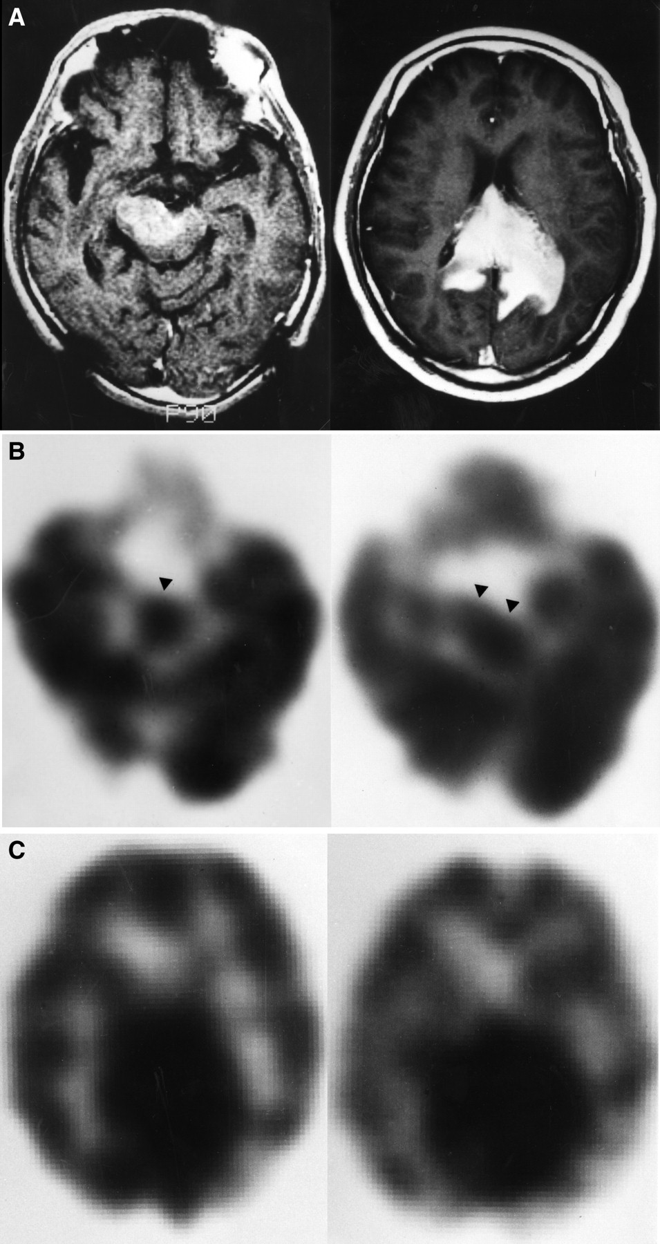
A 69-y-old man with CNS lymphoma (patient 2). (A) T1-weighted MR image with gadolinium-diethylenetriaminepentaacetic acid shows homogenous enhancing lesions in midbrain and corpus callosum. (B) Midbrain tumor shows normal and high accumulation (arrowheads) on early (left) and delayed (right) SPECT images, respectively. (C) Early (left) and delayed (right) 123I-IMP SPECT images reveal increased uptake at corpus callosum.

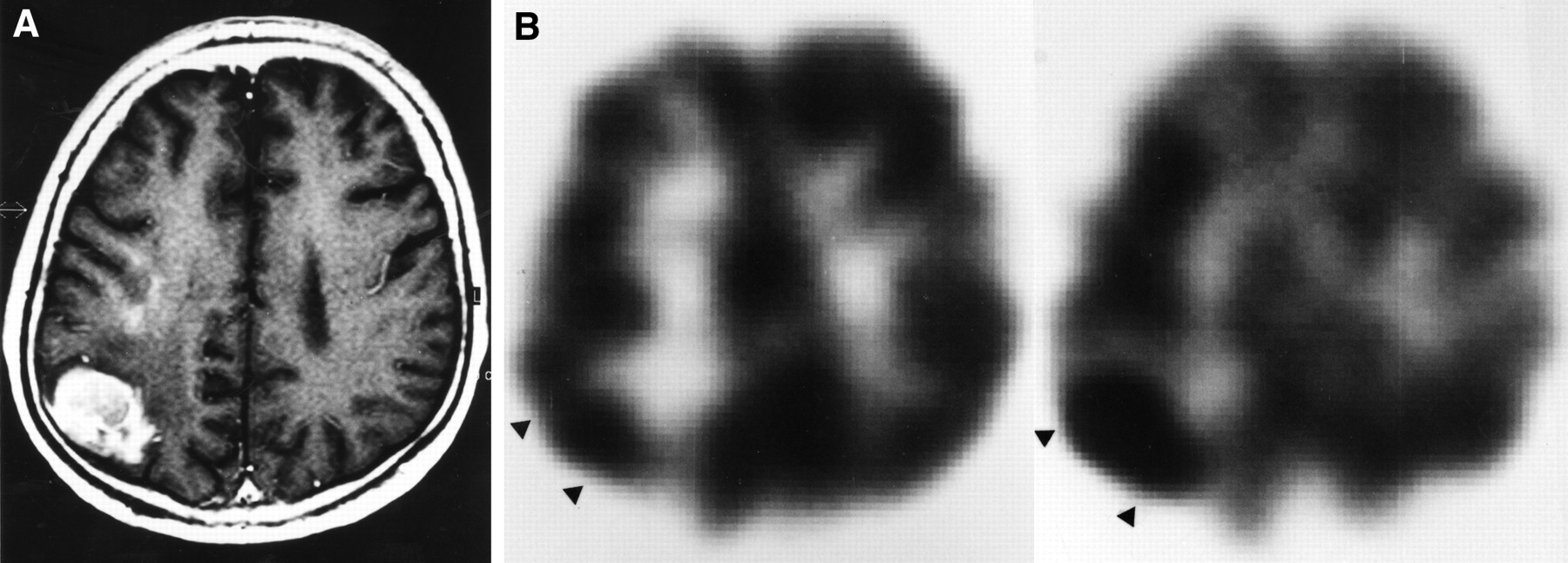
A 52-y-old man with CNS lymphoma (patient 11). (A) T1-weighted MR image with gadolinium-diethylenetriaminepentaacetic acid shows heterogeneously enhancing mass in right parieto-occipital area. Mass is difficult to distinguish from high-grade glioma on MR image only. (B) 123I-IMP SPECT images show normal and increased accumulation (arrowheads) corresponding to tumor on early (left) and delayed (right) images, respectively.

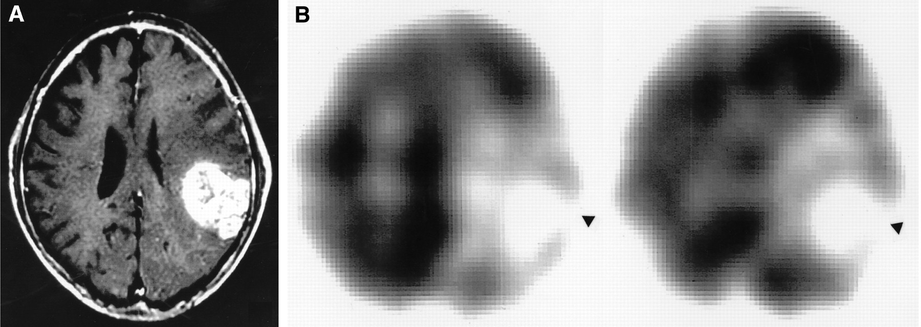
A 72-y-old man with glioblastoma. (A) T1-weighted MR image with gadolinium-diethylenetriaminepentaacetic acid shows heterogeneously enhancing mass in left parietal region. (B) 123I-IMP SPECT images show defect (arrowheads) corresponding to tumor on both early (left) and delayed (right) images. T/Ns on early and delayed images are 0.23 and 0.21, respectively. T/Cs on early and delayed images are 0.18 and 0.20, respectively.

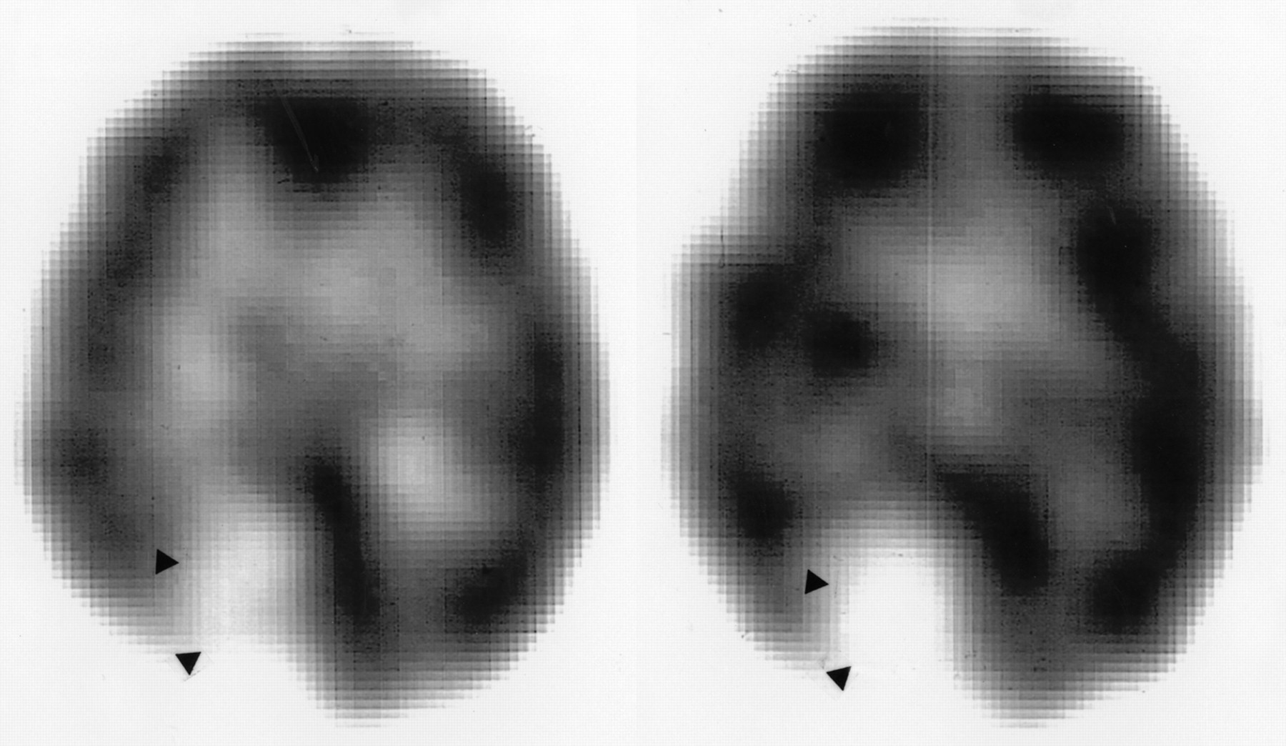
A 71-y-old woman with right parasagittal meningioma. 123I-IMP SPECT images show low accumulation and defect (arrowheads) corresponding to tumor on early (left) and delayed (right) images, respectively. T/Ns on early and delayed images are 0.53 and 0.48, respectively. T/Cs on early and delayed images are 0.18 and 0.14, respectively.
Clinical Features and 123I-IMP SPECT Results for Patients with Non-Hodgkin's Lymphoma of Central Nervous System
The T/Ns and T/Cs of CNS lymphomas, gliomas, and meningiomas are shown in Table 2. On early images, the T/Ns and T/Cs of CNS lymphomas were significantly different from those of gliomas (P < 0.05) but not from those of meningiomas (P = 0.10 and 0.59, respectively). On delayed images, the T/Ns and T/Cs of CNS lymphomas were significantly different from both gliomas and meningiomas (P < 0.0005).
Results of Semiquantitative Analysis of CNS Lymphoma, Gliomas, and Meningioma
DISCUSSION
123I-IMP SPECT and Brain Tumors
123I-IMP is a lipid-soluble radiopharmaceutical agent whose early distribution pattern reflects regional cerebral blood flow (5), because IMP shows high first-pass uptake by brain tissues with nonspecific amine binding sites and reveals slow washout. The possibility has been suggested that brain tumors are visualized as defects on 123I-IMP SPECT because of a lack of binding sites for IMP or because of the absence of the intracellular metabolic pathway for IMP (9,11). Although some brain tumors with high IMP uptake in the early phase have been reported (12–14,16), dynamic SPECT scan studies have revealed that these accumulations resulted from intravascular retention of 123I-IMP (16).
Delayed SPECT images may reflect the distribution of hydrophilic metabolites of IMP resulting from further oxidation of the aliphatic side chain (24). The clinical significance of delayed images remains uncertain (7,10). To our knowledge, only 8 patients with brain tumors showing high uptake in the delayed phase have been described in the literature. We summarize these patients in Table 3. Although 1 patient with malignant astrocytoma was reported (16), this case was exceptional in the light of previous studies (10,11). Moretti et al. (25) showed that normal astrocytes, unlike astrocytoma cells, take up IMP. Nakano et al. (16) reported 1 case of metastatic brain tumor but did not describe its histology or tissue of origin.
Patients Previously Reported with Increased 123I-IMP Uptake on Delayed SPECT
Diagnosing CNS Lymphoma and 123I-IMP SPECT
Although MRI and CT are useful in detecting mass lesions, including CNS lymphoma, CNS lymphoma is difficult to distinguish from other CNS disorders (3). Ruiz et al. (26) reported the value of 201Tl brain SPECT for differentiating CNS lymphoma from Toxoplasma encephalitis. Hoffman et al. (27) suggested that FDG PET may have a potential role in differentiating lymphoma from infectious CNS disorders. Both 201Tl SPECT and FDG PET may be useful diagnostic tools for CNS lymphoma. FDG PET can evaluate lesions less than 1 cm in diameter with high spatial resolution. The role of 201Tl SPECT and FDG PET has been limited to distinguishing between neoplastic and non-neoplastic abnormalities in AIDS patients. PET is not widely available because of its high cost. Biopsy, especially stereotactic biopsy as a less invasive technique, is essential in cases that are confusing. In some cases, biopsy may fail to confirm the diagnosis because of reactive glial tissue, infiltrating T cell lymphocytes, and necrotic tissue in CNS lymphomas. For these reasons, another modality is necessary to confirm the diagnosis. With early diagnosis of CNS lymphoma using noninvasive techniques, whole-brain radiation therapy can be initiated in hopes of improving survival and decreasing hospitalization time.
Nakano et al. (16), Ohkawa et al. (18), and Yoshizawa et al. (20) each reported a case of primary CNS lymphoma that revealed high uptake on delayed images. Kitanaka et al. (15) reported a case of metastatic CNS lymphoma showing high accumulation on delayed SPECT images. Those authors suggested that the unusual increased accumulation of IMP on delayed images might be specific for CNS lymphomas. In that study of 19 brain tumors, CNS lymphoma was the only histologic type showing high uptake on delayed images using 123I-IMP SPECT. An increase in the concentration of nonspecific amine receptors on CNS lymphoma cells has been suggested as a mechanism for the increased uptake of 123I-IMP (15,18). This hypothesis has yet to be confirmed because few patients with CNS lymphoma have been studied by 123I-IMP SPECT and because 123I-IMP can no longer be obtained in some countries. 99mTc-hexamethylpropyleneamine oxime (HMPAO) and 99mTc-ethyl cysteinate dimer (ECD) are other radiopharmaceuticals used for measuring regional cerebral blood flow, but their uptake in CNS lymphoma has not been examined. The mechanism of HMPAO and ECD uptake into brain differs from that of IMP. The former is related to the intracellular content of glutathione (28), whereas the latter is associated with intracellular and membranous esterase activity (29).
Clinical Significance of 123I-IMP SPECT for Diagnosing CNS Lymphoma
We assessed the findings of 123I-IMP SPECT in 18 CNS lymphomas through visual and semiquantitative analyses. Our results indicate that both visual analysis and semiquantitative evaluation with T/N and T/C are potentially useful in distinguishing CNS lymphoma from other types of brain tumors. CNS lymphomas more than 3.0 cm3 in volume were detected as obvious foci of high accumulation, including brain stem tumors and intraventricular tumors. In tumors less than 3 cm3 in volume, the contrast was not sufficient to detect the tumor as a definite area of high accumulation. This finding might have been caused in part by uptake of IMP into normal brain as well as into CNS lymphoma and by the low spatial resolution of our imaging system. In evaluating tumors less than 3 cm3 in volume, semiquantitative analysis is recommended to enhance efficacy.
Distinguishing between CNS lymphoma and CNS toxoplasmosis in AIDS patients is a clinical challenge. Our study had no HIV-positive patients. Future studies should evaluate the role of 123I-IMP SPECT in patients with AIDS.
CONCLUSION
Delayed 123I-IMP SPECT images of patients with CNS lymphoma showed specific accumulations corresponding to the actual tumor mass. This finding confirmed the clinical usefulness of 123I-IMP SPECT in diagnosing CNS lymphoma and in differentiating CNS lymphoma from other types of brain tumors. Future studies will be valuable for evaluating the role of 123I-IMP SPECT in detecting CNS lymphoma in AIDS patients.
Acknowledgments
The authors thank Akio Komatsu, Susumu Yao, Masato Honda, and Akihiko Matsumura of The Radiological Center of Shimane Medical University for excellent technical assistance.
Footnotes
Received Oct. 29, 1999; revision accepted Mar. 8, 2000.
For correspondence or reprints contact: Yasuhiko Akiyama, MD, PhD, 89-1 Enya-cho, Izumo Shimane 693-8501, Japan.
REFERENCES
In this issue


Jump to section
Related Articles
Cited By...
- No citing articles found.