


Abstract
Many studies suggest that changes in the uptake of the glucose analog FDG after therapy, compared with pretreatment uptake, predicts tumor response to therapy. However, clinical interpretation is compromised by a limited understanding of the effect of therapy on FDG and 2-deoxy-d-glucose (DG) uptake at the tumor cell level. Methods: Uptake of 2-deoxy-d-[1-3H]glucose (3H-DG) by SW620 colonic tumor cells was measured before and 8, 16, 24, and 48 h after treatment with the novel platinum drug oxaliplatin and the novel thymidylate synthase inhibitor Tomudex. Glucose transport was determined by measuring the initial rate of uptake of the nearly nonmetabolized glucose analog 3-O-methyl-d-[1-3H]glucose (3H-OMG). The effect of these drugs on cell cycle kinetics was determined using flow cytometry. Results: Treatment of SW620 cells with oxaliplatin was found to decrease uptake of 3H-DG after up to 24 h, but uptake returned to control levels after longer treatment. The initial decrease in 3H-DG incorporation was associated with a lower rate of glucose transport. Treatment of cells with Tomudex induced an increase in 3H-DG uptake that depended on treatment duration. Both glucose transport and the volume of distribution of 3H-OMG were higher in Tomudex-treated cells than in control cells. Flow cytometry showed that oxaliplatin induced a G2 and M arrest, whereas a buildup of cells in the S phase was associated with Tomudex treatment. Both treatments induced apoptosis in SW620 cells. Conclusion: Changes in uptake of DG by SW620 colonic tumor cells responding to therapy is specific to the drug type. Modulation of glucose transport was associated with changes in 3H-DG uptake.
Enhanced glucose use by tumor cells through glycolysis, identified by Warburg (1), is a characteristic exploited in cancer imaging with FDG PET. The glucose analogs 2-deoxy-d-glucose (DG) and FDG, like glucose, enter cells and are phosphorylated to DG and FDG phosphates. However, because of the unsuitability of the phosphates as a substrate for glucose phosphate isomerase and the low levels of glucose-6-phosphatase in tumors (2), DG and FDG phosphates undergo little further metabolism.
FDG PET has been shown to be useful in tumor imaging (3). One of the conclusions of a collective European PET oncology workshop (4) was that changes in the uptake of FDG after 1 or 2 cycles of chemotherapy, compared with pretreatment uptake, can predict clinical outcome. Chemotherapy-induced changes in the uptake of FDG have been shown to predict response in many tumor types (5).
Although decreased tumor FDG uptake has been shown to be associated with response, its significance at the tumor cell level is not known. A few in vitro studies have shown that treatment of tumor cells with 2 types of anticancer agents, platinum drugs (6,7) and DNA chain-terminators (8,9), influences the uptake of DG and FDG.
In this study, we examined the effect of the novel platinum agent oxaliplatin on DG uptake by SW620 colonic tumor cells. Controversy exists about whether the rate-limiting step of DG uptake is transport (10), phosphorylation (11), or dephosphorylation of DG phosphate (2). In contrast to previous studies (6,7) on platinum-drug–induced changes in DG uptake, our study determined the effect of oxaliplatin on glucose transport. We also examined the effect of a further class of anticancer drug, the thymidylate synthase inhibitor Tomudex (AstraZeneca, London, UK), on DG uptake and glucose transport. In addition, flow cytometry studies were performed to determine the relationship between cell cycle distribution and DG uptake.
MATERIALS AND METHODS
Cell Culture and Materials
SW620 cells were maintained in Dulbecco's modified Eagle's medium supplemented with penicillin (5000 IU/100 mL) and streptomycin (5000 μg/100 mL) and 10% fetal bovine serum in a humidified 5% CO2:95% air incubator at 37°C. The 2 chemotherapy agents, Tomudex and oxaliplatin, were obtained from AstraZenica and Sanofi-Synthelabo Inc. (New York, NY), respectively. The compounds 2-deoxy-d-[1-3H]glucose (3H-DG) (326 GBq/mmol) and 3-O-methyl-d-[1-3H]glucose (3H-OMG) (167 GBq/mmol) were obtained from Amersham International (Amersham, UK) and were used at a 1:20 working dilution. Cell viability was determined using trypan blue dye exclusion.
Cell Treatment
Confluent cells in a 75 cm2 flask were trypsinized and seeded into 25 cm2 flasks at 0.3 × 10⁶ cells. Four days later, control and treated flasks were set up in triplicate. Treatment times were 8, 17, 25, and 48 h. The drug doses used—15 nmol/L for Tomudex and 2 μmol/L for oxaliplatin—were 10 times the inhibitory concentration of 50% for each drug (H.E.R. Ford, oral communication, August 1999). To ensure that the lower cell density associated with treated cells did not influence uptake of 3H-DG, flasks seeded with 0.6 × 10⁶ cells were also used for 48-h treatments so that the cell density in treated flasks was not greatly different from that in control flasks at the time of the uptake measurements. Similar results for 3H-DG uptake were obtained with both 0.3 × 10⁶ and 0.6 × 10⁶ cell densities. Medium levels were adjusted accordingly to maintain similar cell-to-medium ratios.
Uptake of 3H-DG
Thirty minutes before the addition of isotopes, the cell medium was renewed. Afterward, 75 μL diluted 3H-DG (final concentration, 37 MBq/mL) were added to the cells, which were then returned to the incubator. After 30 min, the incubation medium was discarded and the cells were washed 5 times with 10 mL phosphate-buffered saline (PBS). The cells were then trypsinized by the addition of 1 mL trypsin solution. After neutralizing the trypsin by the addition of 1 mL Dulbecco's modified Eagle's medium plus 10% fetal bovine serum, the cells were worked gently up and down a 1-mL pipette tip to disaggregate them. One milliliter of cells was then added to 5 mL scintillant in a counting vial for counting of 3H. The remaining cells were spun down and resuspended in 200 μL PBS. The viable cell number was determined on 20 μL cell suspension, and the remaining cells were fixed for cell cycle analysis by the addition of ice-cold ethanol to a concentration of 70%.
Glucose Transport
Glucose transport was determined by measuring the initial rate of uptake of the glucose analog 3H-OMG. 3H-OMG is a substrate for glucose transporters but is only slowly metabolized; thus, its initial uptake is considered a measure of glucose transport (12). This uptake was measured in the presence of glucose, because glucose deprivation has been shown to induce apoptosis in some cell types (13,14). After the cells had been preincubated for 45 min in fresh medium, the flasks were briefly inverted so that the medium was no longer in contact with the cells. Seventy-five microliters (140 kBq) of 3H-OMG were added to the medium, and the flask was gently rocked to facilitate mixing. The flasks were then rapidly reinverted to bring the medium back into contact with the cells and initiate incubation with the 3H-OMG. For incubation periods of 30 s or longer, the flasks were replaced in the incubator to prevent appreciable decreases in temperature. At the end of the incubation period, 50 mL ice-cold (0°C) PBS were rapidly (in <0.5 s) poured into the flask to stop further 3H-OMG uptake. The flasks were given 2 further rapid washes with ice-cold PBS. The control and treated flasks were interleaved during 3H-OMG uptake determinations to ensure that both were handled identically. The cells were then trypsinized and treated as for the 3H-DG uptake studies. Each time point was performed in triplicate with the exception of the oxaliplatin-treated cells incubated for 5 s with 3H-OMG, for which each time point was performed in sextuplicate.
Flow Cytometry
Fixed cells that had been stored at −20°C were spun down and resuspended in 0.8 mL PBS. Clumped cells were disaggregated by passage through a 25-gauge needle. RNase (100 μL of a 1 mg/1 mL solution) and 100 μL propidium iodide (0.4 mg/mL) were then added, and the suspended cells were incubated at 37°C for 30 min and then overnight at 4°C. Analysis was then performed on an EPICS Elite ESP flow cytometer (Beckman Coulter UK Ltd., Bucks, UK) using an argon-ion laser tuned to 488 nm and measuring forward and orthogonal light scatter and red fluorescence. G1 and sub-G1 nuclei were sorted from samples exhibiting sub-G1 peaks and then analyzed under fluorescent light microscopy for discrete DNA fragmentation.
Statistics
Means are quoted ±SD and were compared using the Student t test. Numbers of replicates are shown on the figure captions.
RESULTS
Cell Number
Figure 1 shows the effect of treatment of SW620 cells with 15 nmol/L Tomudex and 2 μmol/L oxaliplatin on cell number. Tomudex treatment resulted in significantly lower cell numbers compared with control levels. The cell number was 76% (t6 = 7.44, P < 0.002) of control levels in 17-h treated flasks and decreased to 52% (t6 = 9.31, P < 0.001) after 48 h. Treatment of cells with oxaliplatin also resulted in significantly lower cell numbers compared with control levels at the 48-h time point (t6 = 14.1, P < 0.001), at which treated cells were 65% of control levels. After 25 h, the cell number was insignificantly lower (t6 = 1.6 [not significant (NS)]). Cell viability was more than 97% in all cases.
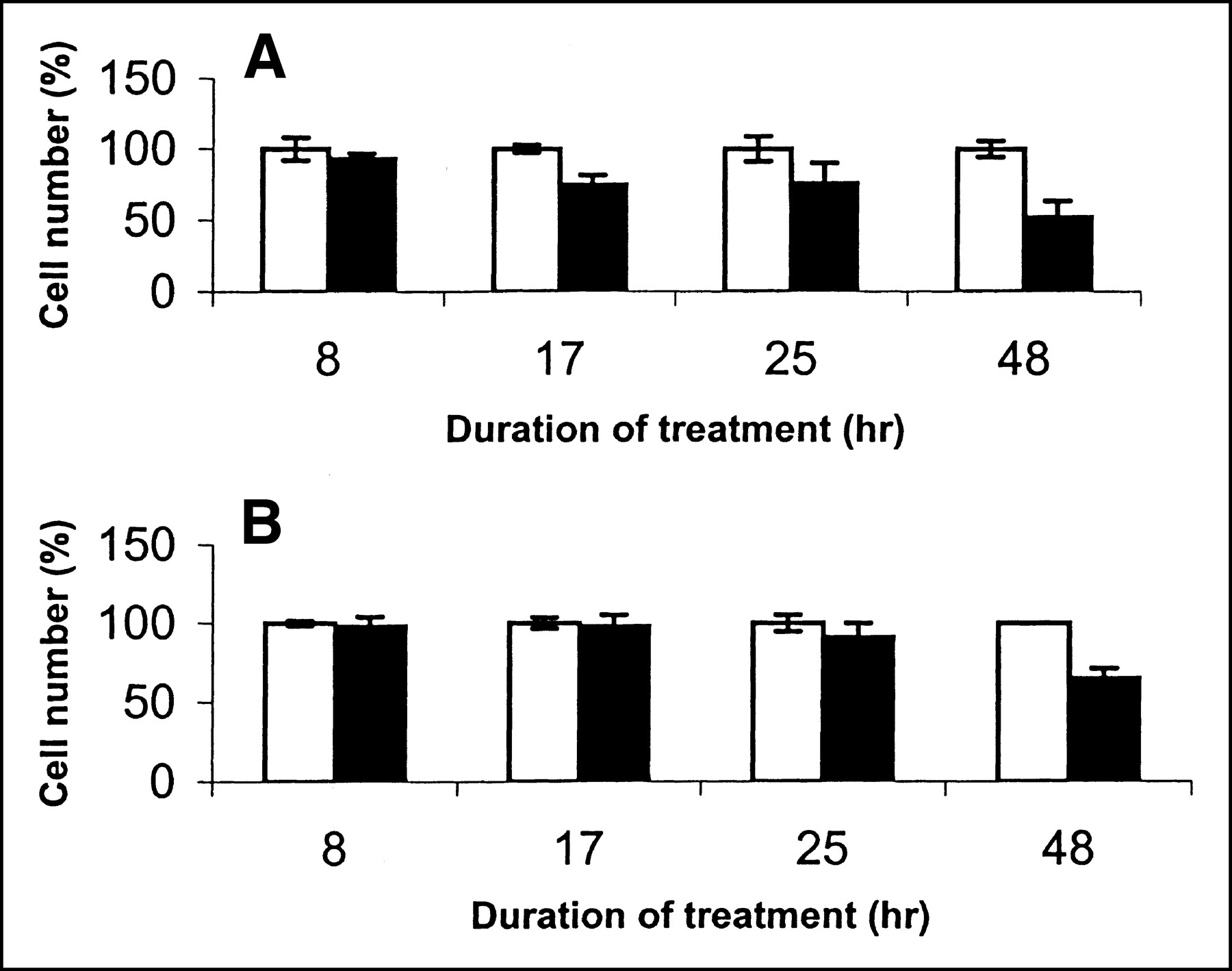
Adherent cell numbers at 8, 17, 25, and 48 h in control flasks (white) and flasks treated (black) with Tomudex (A) and oxaliplatin (B) (n = 3 for control and treated at each time point).
Flow Cytometry
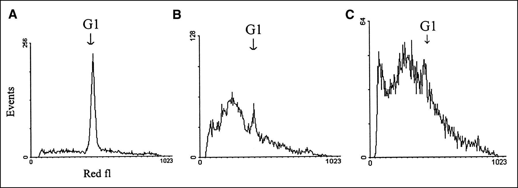
Figures 2 and 3 show the sequence of DNA histograms during treatment of SW620 cells with Tomudex and oxaliplatin, respectively. With increasing duration of exposure, Tomudex increased the proportion of cells in the early to mid S phase. Oxaliplatin treatment initially induced an increase of cells in the S phase, but after longer treatment a G2 block was observed.

DNA histograms, including percentage of cells in each part of cell cycle, from control flasks (A) and flasks treated with Tomudex for 17 (B), 25 (C), and 48 h (D). fl = flask.

DNA histograms, including percentage of cells in each part of cell cycle, from control flasks (A) and flasks treated with oxaliplatin for 17 (B), 25 (C), and 48 h (D). fl = flask.
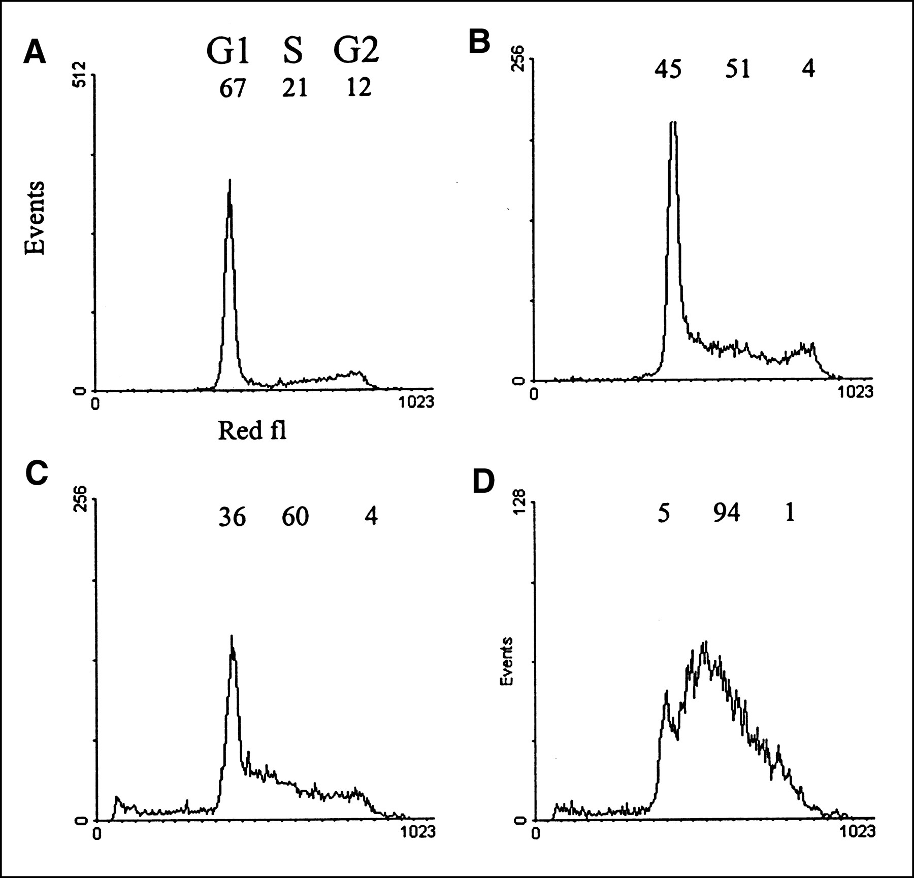
Cells that had become detached after 48 h of treatment with their respective drugs and untreated suspended SW620 cells were examined for apoptosis. Sub-G1 peaks, absent from the control cells, were clearly present (Fig. 4) in samples taken from medium after 48 h of treatment with both Tomudex and oxaliplatin. This finding was consistent with apoptosis and was confirmed using fluorescent-light microscopy after sorting G1 and sub-G1 nuclei. The G1 cells were intact, whereas the sub-G1 cells appeared as fragmented nuclei.
DNA histograms from detached cells untreated (A) and after 48 h treatment with Tomudex (B) and oxaliplatin (C). fl = flask.
3H-DG Uptake
Figure 5 shows the effect of treatment of SW620 cells with Tomudex and oxaliplatin on the uptake of 3H-DG per 106 cells. After cells were treated 8 h with Tomudex, 3H-DG uptake rose slightly but insignificantly (t6 = 1.69, NS) compared with control levels. Increased uptake compared with control levels occurred at 17 h (t6 = 3.83, P < 0.02), 25 h (t6 = 3.03, P < 0.05), and 48 h (t6 = 7.22, P < 0.001) after treatment. Treatment of cells with oxaliplatin resulted in a significant decrease in 3H-DG uptake after 8 h (t6 = 3.78, P < 0.05), with a further decrease after 17 h (t6 = 8.81, P < 0.001). 3H-DG uptake was also lower than control levels at 25 h (t6 = 3.4, P < 0.05) but returned to control levels after 48 h.

DG incorporation at 8, 17, 25, and 48 h by control SW620 cells (white) and SW620 cells treated (black) with Tomudex (A) and oxaliplatin (B) (n = 3 for control and treated at 8, 17, and 25 h; n = 6 for control and treated at 48 h).
3H-OMG Uptake
Figure 6 shows the uptake of 3H-OMG by control cells after 3, 5, 7, 15, 30, and 300 s of incubation with this glucose analog. Uptake of this compound was found to be linear for approximately the first 7–10 s. Incubations of 7 s or less with 3H-OMG were therefore considered measures of initial uptake rate and used as an estimate of glucose transport. Equilibrium was shown to occur after approximately 30 s of incubation.

Time course of OMG uptake by control SW620 cells (n = 3 for each time point).
Figure 7A shows the effect of treatment of SW620 cells with Tomudex for 25 h on uptake of 3H-OMG. Uptake of 3H-OMG was found to be significantly higher than control levels after 7 (t6 = 4.39, P < 0.02), 15 (t6 = 5.40, P < 0.01), and 300 (t6 = 2.74, P < 0.05) s of incubation. After 30 s, uptake was higher, although not significantly so.
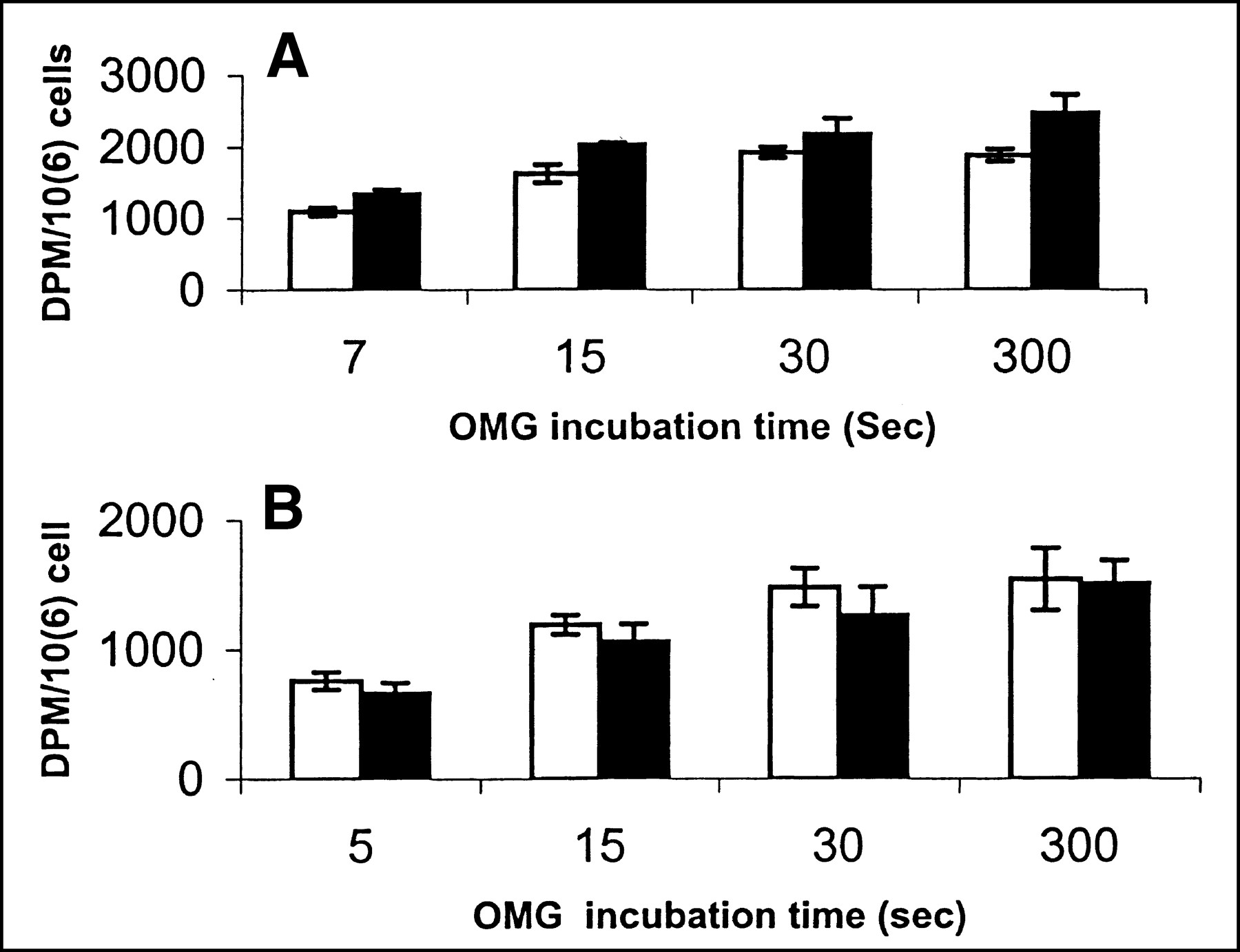
OMG uptake by control cells (white) and cells treated (black) for 25 h with Tomudex (A) and oxaliplatin (B) after various incubation periods with OMG (n = 3 or more).
Figure 7B shows the effect of treatment of SW620 cells with oxaliplatin for 25 h on uptake of 3H-OMG. Significantly lower uptake—approximately 13%—of 3H-OMG was seen at the 5-s incubation time (t12 = 2.30, P < 0.05). Although the uptake curve for treated cells was lower than for control cells at 15 and 30 s, uptake of 3H-OMG was not significantly different at these time points. After 300 s of incubation, the 2 curves converged.
Uptake of 3H-OMG after 5 s of incubation with 3H-OMG was found to be 66% higher (t = 10.8, P < 0.001) by cells treated for 48 h with Tomudex (1559 ± 74, n = 4) than by control cells (939 ± 97, n = 6), corresponding with the trend in 3H-DG uptake. After 48 h of incubation with oxaliplatin, 3H-OMG uptake (1159 ± 201, n = 6) during a 5-s incubation was found to be 23% higher (t = 2.56, P < 0.05) than control levels (941 ± 154, n = 12).
The proportion of uptake after 5 s as a consequence of passive diffusion was determined in control cells after preincubation for 10 min with the facilitative glucose transport inhibitor cytochalasin B at a high concentration (20 μmol/L). Incubation with this inhibitor reduced 3H-OMG uptake from 736 ± 99 (n = 6) to 266 ± 25 dpm per 106 cells (n = 6), indicating that the majority (64%) of 3H-OMG transport occurred through facilitative glucose transport.
DISCUSSION
Treatment of SW620 colonic tumor cells with Tomudex and oxaliplatin was associated with changes in cell number, cell cycle distribution, incorporation of 3H-DG, and initial uptake of 3H-OMG. Both drugs ultimately induced apoptosis in this cell line. Tomudex induced a treatment duration–related increase in cells in the S phase consistent with a block in the early or mid S phase. The accumulation of cells in the S phase was paralleled by increased uptake of 3H-DG. Oxaliplatin treatment was initially associated with a buildup of cells in the S phase, but after 48 h a G2 accumulation was clearly apparent. This drug initially induced a decrease in 3H-DG incorporation, but after longer treatments uptake returned to nontreatment levels.
Both oxaliplatin and cisplatin induce DNA adducts and DNA cross-links (15) blocking DNA replication and inducing cell cycle arrest. Studies have also shown that in common with cisplatin (16), oxaliplatin induces cell-cycle arrest in the G2 and M phases (17). Haberkorn et al. (6) examined the effect of cisplatin and also of 2 novel platinum compounds linked to phosphonic acid, that is, cis-diamine-[nitrilobis(methylphosphonato)(2-)O1,N1]platinum(II) (AMDP) and cis-cyclohexane-1,2-diamine-[nitriloTris(methylphosphonato)(2-)-O1,N1]platinum(II) (DADP), on the uptake of FDG by 2 cell lines derived from a metastasis of a rat osteocarcinoma. AMDP induced a buildup of cells in G2 after a 4-h treatment and a block in S after longer treatments. AMDP-induced changes in FDG varied and depended on cell line. Initially, 4 h of treatment were found to decrease the uptake of FDG in 1 cell line when determined 1 h after treatment. However, in common with this study, uptake of FDG then increased to approximately untreated levels after longer treatment. Treatment with cisplatin resulted in a buildup of cells in G2. Both cisplatin and DADP caused an increase in FDG uptake 24 h after the end of treatment with cisplatin and DADP. This increase was particularly pronounced after DADP treatment, which appeared not to induce significant cell cycle changes.
These results show that platinum-drug–induced modifications of DG uptake by cancer cell lines depends on both cell type and platinum compound and does not appear to be related to cell cycle. By examining the initial uptake of 3H-OMG by SW620 cells 25 h after exposure to oxaliplatin, this study found a decrease in the transport of glucose that was comparable with the lower rate of uptake of DG by cells treated with this drug. A study showed that treatment of tumor cells with platinum compounds inhibited uptake of amino acids (18). In addition to the DNA-damaging effects of platinum drugs, sulfhydryl groups in proteins have been shown to be direct targets of these compounds (19). Sulfhydryl groups are found within the glucose transporter-1 molecule (20); thus, treatment of cells with oxaliplatin may initially decrease 3H-OMG and 3H-DG uptake by SW620 cells by binding to such groups on glucose transporters. The increased rate of glucose transport after 48 h of treatment may be attributed to new glucose transporter protein synthesis in response to decreased glucose uptake resulting from glucose transporter inactivation.
A further possible mechanism for oxaliplatin-induced modification of glucose uptake may be proposed from the finding that cisplatin induces phosphorylation of tyrosine in several proteins (21). Phosphorylation of glucose transporters changes their substrate affinity and, thus, is associated with modulation of hexose transport.
Treatment of SW620 cells with Tomudex induced a buildup of cells in the S phase and increased DG uptake in a manner dependent on treatment duration. Two studies using agents that, in common with Tomudex, induce arrest in the S phase, but through a different mechanism, have shown an association with at least an initial increase in FDG uptake (8,9). In the first study, a time- and dose-dependent increase in FDG uptake by rat prostate adenocarcinoma cells treated with the deoxycytidine analog gemcitabine was observed (9). The second study found that sensitized Morris hepatoma cells treated with the DNA chain terminator ganciclovir exhibited increased uptake of FDG after 24 h (8), declining to untreated levels after 48 h of treatment. 3H-OMG uptake during a 10-min incubation after treatment with ganciclovir was also measured and found to be increased at both time points. Uptake of 3H-OMG during 10 min will probably have reached equilibrium, so Haberkorn et al. (8) would have been measuring the volume of distribution of 3H-OMG. In this study, both the initial uptake rate of 3H-OMG, measured after 5 s, and its cell concentration at equilibrium were determined and found to be increased in cells treated for 25 h with Tomudex. The increase in volume of distribution may be caused by changes in the cell cycle distribution. Such changes have been shown to be associated with changes in mean cell volume (22). Luc et al. (22) showed that the mean cell volume of isolated tumor cells in G1 and G0 was approximately 65% of that of populations of unseparated cells. Larger cells are more likely to have higher volumes of distribution. Having a larger plasma membrane surface area, larger cells may also possess greater numbers of glucose transporters and exhibit faster initial 3H-OMG uptake.
Both Tomudex and oxaliplatin were found to induce apoptosis, and this was apparent in cells that had become detached after 48 h of treatment. Uptake of 3H-DG by cells treated by these drugs may be influenced by the onset of apoptosis. A study showed that the induction of apoptosis in Jurkat cells by CD95 ligation was accompanied by an early decrease in glucose transport as a consequence of reduced affinity of the glucose transporter (23). The initial decrease in glucose transport associated with treatment of SW620 cells with oxaliplatin may be caused by a similar mechanism.
The corresponding change in glucose transport with DG incorporation into SW620 cells after treatment with either oxaliplatin or Tomudex suggests that in this cell type, transport, rather than glucose phosphorylation by hexokinase, is the rate-limiting step in the early part of the glycolytic pathway. This finding agrees with previous findings (10).
In vivo studies have generally shown that response to therapy is associated with a decreased uptake of FDG (5). However, some clinical studies have performed FDG scanning weeks or even months after the completion of therapy, when tumor cell response to the drug may no longer be evident. Further, modulations in blood flow and inflammatory response may contribute to therapy-induced changes in uptake of FDG by solid tumors. In this study, we saw that responding tumor cells can show increased or decreased uptake of DG, at least in the short term, depending on the type of drug therapy. Therefore, an increase in uptake of FDG by tumors after some forms of chemotherapy may not necessarily indicate a lack of response.
CONCLUSION
We examined the uptake of 3H-DG by SW620 cells after treatment with 2 novel chemotherapy drugs for colonic cancer. The uptake patterns for the 2 drugs were different, with Tomudex showing a treatment-duration–related increase in uptake associated with increased glucose transport. Oxaliplatin showed a more complex pattern of DG uptake, presumably reflecting the variety of cellular effects induced by this drug. Changes in glucose transport corresponded with the increase in 3H-DG uptake exhibited by Tomudex-treated cells and the initial decrease in 3H-DG uptake shown by oxaliplatin-treated cells.
Acknowledgments
The authors acknowledge funding from Cancer Research Campaign grant SP1780/0103 and National Institutes of Health grant CA 62557-02.
Footnotes
Received Nov. 5, 1999; revision accepted Mar. 28, 2000.
For correspondence or reprints contact: Timothy A.D. Smith, PhD, MRC Cyclotron Unit, Hammersmith Hospital, Du Canc Rd., London W120NN, UK.
REFERENCES
In this issue


Jump to section
Related Articles
Cited By...
- Colorectal Tumor Cells Treated with 5-FU, Oxaliplatin, Irinotecan, and Cetuximab Exhibit Changes in 18F-FDG Incorporation Corresponding to Hexokinase Activity and Glucose Transport
- Treatment of Breast Tumor Cells In Vitro with the Mitochondrial Membrane Potential Dissipater Valinomycin Increases 18F-FDG Incorporation
- Quantitation of Small-Animal 124I Activity Distributions Using a Clinical PET/CT Scanner
- 2-[18F]Fluoro-2-deoxyglucose and Glucose Uptake in Malignant Gliomas before and after Radiotherapy: Correlation with Outcome