
Main menu

  • Home
  • Content
    • Current
    • Ahead of print
    • Past Issues
    • JNM Supplement
    • SNMMI Annual Meeting Abstracts
    • Continuing Education
    • JNM Podcasts
  • Subscriptions
    • Subscribers
    • Institutional and Non-member
    • Rates
    • Journal Claims
    • Corporate & Special Sales
  • Authors
    • Submit to JNM
    • Information for Authors
    • Assignment of Copyright
    • AQARA requirements
  • Info
    • Reviewers
    • Permissions
    • Advertisers
  • About
    • About Us
    • Editorial Board
    • Contact Information
  • More
    • Alerts
    • Feedback
    • Help
    • SNMMI Journals
  • SNMMI
    • JNM
    • JNMT
    • SNMMI Journals
    • SNMMI

User menu

  • Subscribe
  • My alerts
  • Log in
  • My Cart

Search

  • Advanced search
Journal of Nuclear Medicine
  • SNMMI
    • JNM
    • JNMT
    • SNMMI Journals
    • SNMMI
  • Subscribe
  • My alerts
  • Log in
  • My Cart
Journal of Nuclear Medicine

Advanced Search

  • Home
  • Content
    • Current
    • Ahead of print
    • Past Issues
    • JNM Supplement
    • SNMMI Annual Meeting Abstracts
    • Continuing Education
    • JNM Podcasts
  • Subscriptions
    • Subscribers
    • Institutional and Non-member
    • Rates
    • Journal Claims
    • Corporate & Special Sales
  • Authors
    • Submit to JNM
    • Information for Authors
    • Assignment of Copyright
    • AQARA requirements
  • Info
    • Reviewers
    • Permissions
    • Advertisers
  • About
    • About Us
    • Editorial Board
    • Contact Information
  • More
    • Alerts
    • Feedback
    • Help
    • SNMMI Journals
  • View or Listen to JNM Podcast
  • Visit JNM on Facebook
  • Join JNM on LinkedIn
  • Follow JNM on Twitter
  • Subscribe to our RSS feeds
OtherClinical Investigations

Evidence of Bilateral Temporal Lobe Involvement in Primary Progressive Aphasia: A SPECT Study

Marie-Hélène Soriani-Lefèvre, Didier Hannequin, Serge Bakchine, Jean-François Ménard, Alain Manrique, Anne Hitzel, Pierre-Olivier Kotzki, Vincent Boudousq and Pierre Vera
Journal of Nuclear Medicine July 2003, 44 (7) 1013-1022;
Marie-Hélène Soriani-Lefèvre
  • Find this author on Google Scholar
  • Find this author on PubMed
  • Search for this author on this site
Didier Hannequin
  • Find this author on Google Scholar
  • Find this author on PubMed
  • Search for this author on this site
Serge Bakchine
  • Find this author on Google Scholar
  • Find this author on PubMed
  • Search for this author on this site
Jean-François Ménard
  • Find this author on Google Scholar
  • Find this author on PubMed
  • Search for this author on this site
Alain Manrique
  • Find this author on Google Scholar
  • Find this author on PubMed
  • Search for this author on this site
Anne Hitzel
  • Find this author on Google Scholar
  • Find this author on PubMed
  • Search for this author on this site
Pierre-Olivier Kotzki
  • Find this author on Google Scholar
  • Find this author on PubMed
  • Search for this author on this site
Vincent Boudousq
  • Find this author on Google Scholar
  • Find this author on PubMed
  • Search for this author on this site
Pierre Vera
  • Find this author on Google Scholar
  • Find this author on PubMed
  • Search for this author on this site
  • Article
  • Figures & Data
  • Info & Metrics
  • PDF
Loading
  1. FIGURE 1.
    • Download figure
    • Open in new tab
    • Download powerpoint
    FIGURE 1.

    Brain systematization. (A) Lateral side. (B) Medial side. Broca = Broca region; Calc = calcarine region; CinA = anterior cingulate region; EF = external frontal region; IF = internal frontal region; Insul = insular region; LobQ = lobulus quadrilatere; LT = lateral temporal region; MT = medial temporal region; Occ = occipital region; OF = orbitofrontal region; P = parietal lobe; PreC = precentral region; PreF = prefrontal region; SM = sensorimotor region; Tha = thalamus; TP = temporal pole; W = Wernicke region.
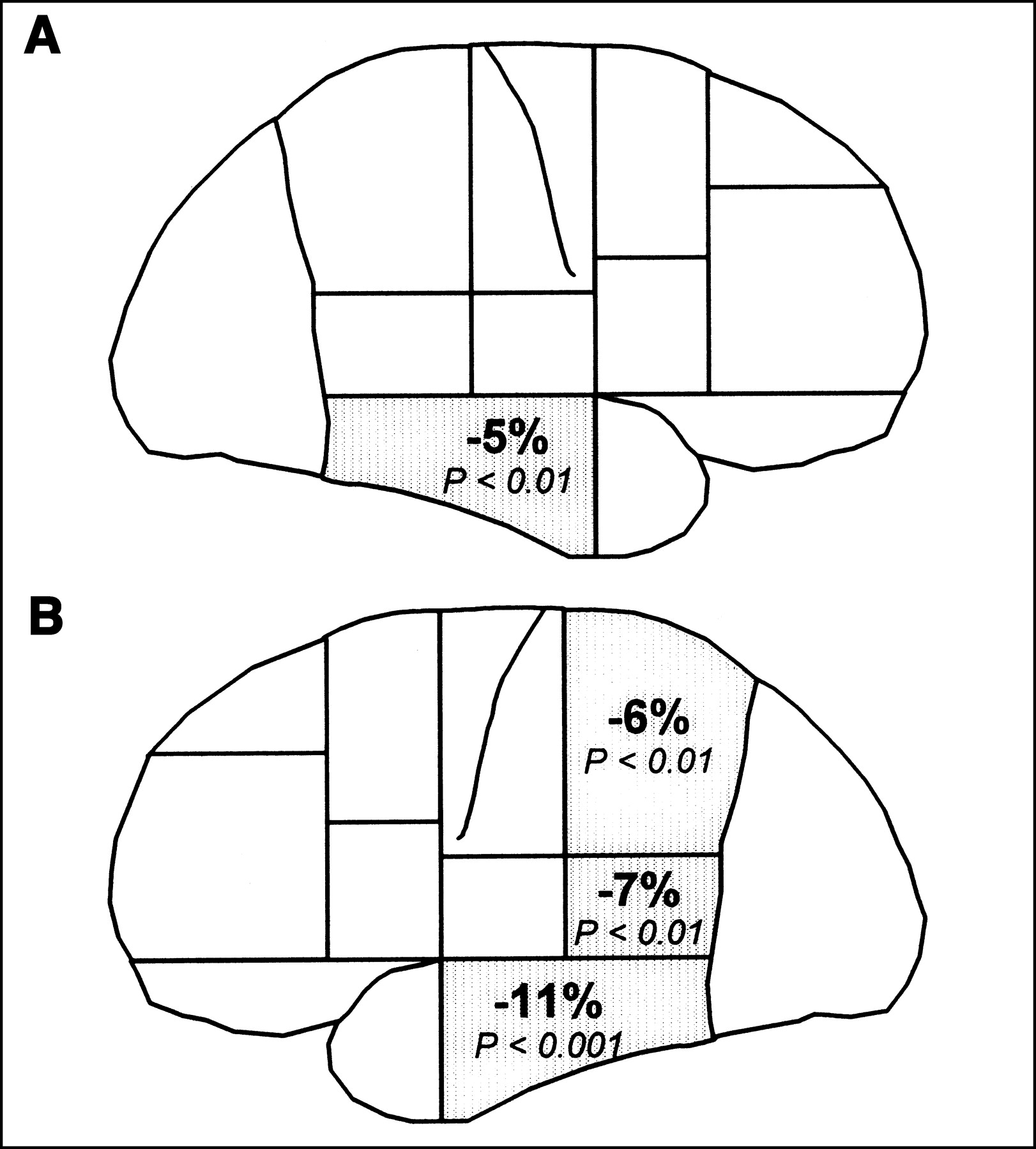
  2. FIGURE 2.
    • Download figure
    • Open in new tab
    • Download powerpoint
    FIGURE 2.

    Percentage of PI decrease in 29 patients vs. 12 control subjects. (A) Right hemisphere. (B) Left hemisphere.
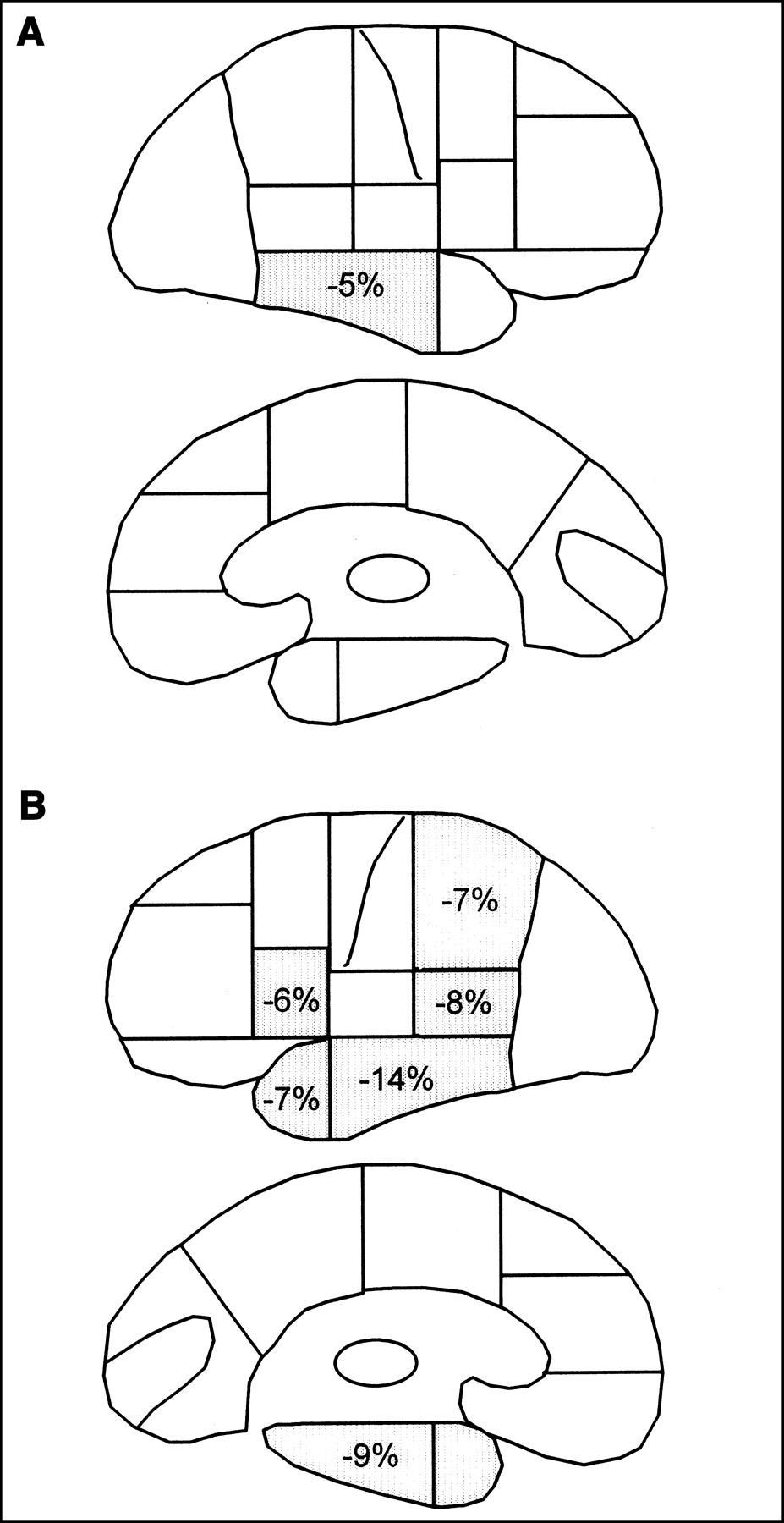
  3. FIGURE 3.
    • Download figure
    • Open in new tab
    • Download powerpoint
    FIGURE 3.

    Comparison of PI in patients with lexicosemantic disorders and control subjects. (A) Right hemisphere, lateral and medial side. (B) Left hemisphere, lateral and medial side.
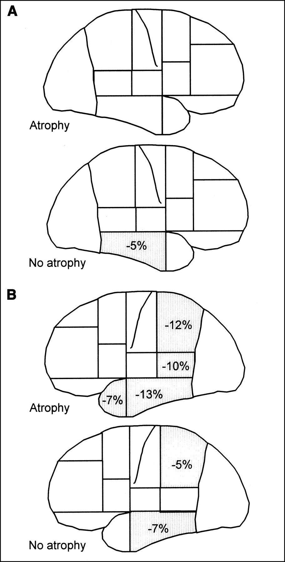
  4. FIGURE 4.
    • Download figure
    • Open in new tab
    • Download powerpoint
    FIGURE 4.

    Comparison of PI in patients with or without atrophy and control subjects. (A) Right hemisphere, atrophic and nonatrophic groups. (B) Left hemisphere, atrophic and nonatrophic groups.

PreviousNext
Back to top

In this issue

Journal of Nuclear Medicine
Vol. 44, Issue 7
July 1, 2003
  • Table of Contents
  • Index by author
Print
Download PDF
Article Alerts
Sign In to Email Alerts with your Email Address
Email Article

Thank you for your interest in spreading the word on Journal of Nuclear Medicine.

NOTE: We only request your email address so that the person you are recommending the page to knows that you wanted them to see it, and that it is not junk mail. We do not capture any email address.

Enter multiple addresses on separate lines or separate them with commas.
Evidence of Bilateral Temporal Lobe Involvement in Primary Progressive Aphasia: A SPECT Study
(Your Name) has sent you a message from Journal of Nuclear Medicine
(Your Name) thought you would like to see the Journal of Nuclear Medicine web site.
Citation Tools
Evidence of Bilateral Temporal Lobe Involvement in Primary Progressive Aphasia: A SPECT Study
Marie-Hélène Soriani-Lefèvre, Didier Hannequin, Serge Bakchine, Jean-François Ménard, Alain Manrique, Anne Hitzel, Pierre-Olivier Kotzki, Vincent Boudousq, Pierre Vera
Journal of Nuclear Medicine Jul 2003, 44 (7) 1013-1022;

Citation Manager Formats

  • BibTeX
  • Bookends
  • EasyBib
  • EndNote (tagged)
  • EndNote 8 (xml)
  • Medlars
  • Mendeley
  • Papers
  • RefWorks Tagged
  • Ref Manager
  • RIS
  • Zotero
Share
Evidence of Bilateral Temporal Lobe Involvement in Primary Progressive Aphasia: A SPECT Study
Marie-Hélène Soriani-Lefèvre, Didier Hannequin, Serge Bakchine, Jean-François Ménard, Alain Manrique, Anne Hitzel, Pierre-Olivier Kotzki, Vincent Boudousq, Pierre Vera
Journal of Nuclear Medicine Jul 2003, 44 (7) 1013-1022;
Twitter logo Facebook logo LinkedIn logo Mendeley logo
  • Tweet Widget
  • Facebook Like
  • Google Plus One
Bookmark this article

Jump to section

  • Article
    • Abstract
    • MATERIALS AND METHODS
    • RESULTS
    • DISCUSSION
    • CONCLUSION
    • Acknowledgments
    • Footnotes
    • REFERENCES
  • Figures & Data
  • Info & Metrics
  • PDF

Related Articles

  • No related articles found.
  • PubMed
  • Google Scholar

Cited By...

  • Natural history of primary progressive aphasia
  • Google Scholar

More in this TOC Section

  • Feasibility of Ultra-Low-Activity 18F-FDG PET/CT Imaging Using a Long–Axial-Field-of-View PET/CT System
  • Cardiac Presynaptic Sympathetic Nervous Function Evaluated by Cardiac PET in Patients with Chronotropic Incompetence Without Heart Failure
  • Validation and Evaluation of a Vendor-Provided Head Motion Correction Algorithm on the uMI Panorama PET/CT System
Show more Clinical Investigations

Similar Articles

SNMMI

© 2025 SNMMI

Powered by HighWire